 Movie
Movie Controller

[English] 日本語
 Yorodumi
- PDB-1ig8: Crystal Structure of Yeast Hexokinase PII with the correct amino ... -
+ Open data

- Basic information
| Entry | Database: PDB / ID: 1ig8 | ||||||
|---|---|---|---|---|---|---|---|
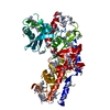
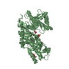
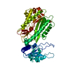
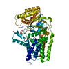
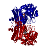
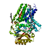
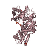
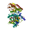
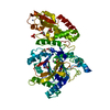
| Title | Crystal Structure of Yeast Hexokinase PII with the correct amino acid sequence | ||||||
 Components Components | hexokinase PII | ||||||
 Keywords Keywords | TRANSFERASE / mixed alpha beta / two domains / cleft | ||||||
| Function / homology |  Function and homology information Function and homology informationSynthesis of GDP-mannose / fructose import across plasma membrane / hexokinase activity / Glycolysis / mannokinase activity / hexokinase / fructokinase activity / glucokinase activity / Regulation of Glucokinase by Glucokinase Regulatory Protein / mannose metabolic process ...Synthesis of GDP-mannose / fructose import across plasma membrane / hexokinase activity / Glycolysis / mannokinase activity / hexokinase / fructokinase activity / glucokinase activity / Regulation of Glucokinase by Glucokinase Regulatory Protein / mannose metabolic process / regulation of transcription by glucose / glucose 6-phosphate metabolic process / D-glucose binding / fructose metabolic process / : / regulation of cell size / negative regulation of apoptotic signaling pathway / Neutrophil degranulation / intracellular glucose homeostasis / glycolytic process / glucose metabolic process / mitochondrial membrane / mitochondrion / ATP binding / nucleus / cytosol Similarity search - Function | ||||||
| Biological species |  | ||||||
| Method |  X-RAY DIFFRACTION / SYNCHROTRON / MOLECULAR REPLACEMENT / Resolution: 2.2 Å X-RAY DIFFRACTION / SYNCHROTRON / MOLECULAR REPLACEMENT / Resolution: 2.2 Å | ||||||
 Authors Authors | Kuser, P.R. / Krauchenco, S. / Antunes, O.A. / Polikarpov, I. | ||||||
 Citation Citation | Journal: J.Biol.Chem. / Year: 2000 Title: The high resolution crystal structure of yeast hexokinase PII with the correct primary sequence provides new insights into its mechanism of action. Authors: Kuser, P.R. / Krauchenco, S. / Antunes, O.A. / Polikarpov, I. #1: Journal: Acta Crystallogr.,Sect.D / Year: 1999Title: Crystallization and Preliminary Crystal Analysis of Yeast Hexokinase PI and PII Authors: Kuser, P.R. / Golubev, A.M. / Polikarpov, I. | ||||||
| History |
|
- Structure visualization
| Structure viewer | Molecule: MolmilJmol/JSmol |
|---|
- Downloads & links
-Download
| PDBx/mmCIF format | 1ig8.cif.gz | 113.7 KB | Display | PDBx/mmCIF format |
|---|---|---|---|---|
| PDB format | pdb1ig8.ent.gz | 87.7 KB | Display | PDB format |
| PDBx/mmJSON format | 1ig8.json.gz | Tree view | PDBx/mmJSON format | |
| Others |  Other downloads Other downloads |
-Validation report
| Arichive directory | https://data.pdbj.org/pub/pdb/validation_reports/ig/1ig8ftp://data.pdbj.org/pub/pdb/validation_reports/ig/1ig8 | HTTPS FTP |
|---|
-Related structure data
| Related structure data | |
|---|---|
| Similar structure data |


-Links
 PDBj
PDBj


- Assembly
| Deposited unit | 
| ||||||||
|---|---|---|---|---|---|---|---|---|---|
| 1 |
| ||||||||
| Unit cell |
|
-Components
| #1: Protein | Mass: 54001.242 Da / Num. of mol.: 1 / Source method: isolated from a natural source / Source: (natural) |
|---|---|
| #2: Chemical | ChemComp-SO4 /   Mass: 96.063 Da / Num. of mol.: 1 / Source method: obtained synthetically / Formula: SO4 Mass: 96.063 Da / Num. of mol.: 1 / Source method: obtained synthetically / Formula: SO4 |
| #3: Water | ChemComp-HOH /  Mass: 18.015 Da / Num. of mol.: 441 / Source method: isolated from a natural source / Formula: H2O Mass: 18.015 Da / Num. of mol.: 441 / Source method: isolated from a natural source / Formula: H2O |
-Experimental details
-Experiment
| Experiment | Method: X-RAY DIFFRACTION / Number of used crystals: 1 |
|---|
- Sample preparation
| Crystal | Density Matthews: 2.76 Å3/Da / Density % sol: 55.41 % | |||||||||||||||
|---|---|---|---|---|---|---|---|---|---|---|---|---|---|---|---|---|
| Crystal grow | Temperature: 298 K / Method: vapor diffusion, hanging drop / pH: 7.5 Details: 2.3-3.2M Ammonium sulphate, pH 7.5, VAPOR DIFFUSION, HANGING DROP, temperature 298.0K | |||||||||||||||
| Crystal grow | *PLUS Method: vapor diffusion, sitting drop | |||||||||||||||
| Components of the solutions | *PLUS
|
-Data collection
| Diffraction | Mean temperature: 277 K |
|---|---|
| Diffraction source | Source: SYNCHROTRON / Site: LNLS  / Beamline: D03B-MX1 / Wavelength: 1.38 Å / Beamline: D03B-MX1 / Wavelength: 1.38 Å |
| Detector | Type: MARRESEARCH / Detector: IMAGE PLATE / Date: Sep 10, 1998 |
| Radiation | Monochromator: Si 111 / Protocol: SINGLE WAVELENGTH / Monochromatic (M) / Laue (L): M / Scattering type: x-ray |
| Radiation wavelength | Wavelength: 1.38 Å / Relative weight: 1 |
| Reflection | Resolution: 2.2→13 Å / Num. obs: 60395 / % possible obs: 95.1 % / Redundancy: 2.12 % / Biso Wilson estimate: 23.13 Å2 / Rsym value: 0.113 |
| Reflection shell | Resolution: 2.2→2.25 Å / Redundancy: 1.82 % / Rmerge(I) obs: 0.42 / Num. unique all: 1914 / % possible all: 96.6 |
| Reflection | *PLUS Rmerge(I) obs: 0.113 |
| Reflection shell | *PLUS % possible obs: 96.6 % |
- Processing
| Software |
| |||||||||||||||||||||||||
|---|---|---|---|---|---|---|---|---|---|---|---|---|---|---|---|---|---|---|---|---|---|---|---|---|---|---|
| Refinement | Method to determine structure: MOLECULAR REPLACEMENT Starting model: P152K mutant of yeast hexokinase Resolution: 2.2→13 Å / SU B: 6.1415 / SU ML: 0.15315 / Isotropic thermal model: Overall / Cross valid method: THROUGHOUT / σ(F): 0 / ESU R: 0.24781 / ESU R Free: 0.22644 / Stereochemistry target values: Engh and Huber
| |||||||||||||||||||||||||
| Displacement parameters | Biso mean: 23.4 Å2 | |||||||||||||||||||||||||
| Refinement step | Cycle: LAST / Resolution: 2.2→13 Å
| |||||||||||||||||||||||||
| Refine LS restraints |
|
