 ムービー
ムービー コントローラー


+ データを開く

- 基本情報
基本情報
| 登録情報 | データベース: PDB / ID: 5w1q | ||||||
|---|---|---|---|---|---|---|---|
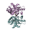
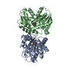
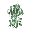
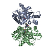
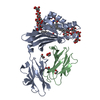
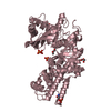
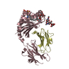
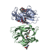
| タイトル | Crystal structure of alternate isoform of glutamate racemase from Helicobacter pylori bound to D-glutamate | ||||||
 要素 要素 | Glutamate racemase | ||||||
 キーワード キーワード | ISOMERASE / racemase / cofactor independent / glutamate | ||||||
| 機能・相同性 |  機能・相同性情報 機能・相同性情報glutamate racemase / glutamate racemase activity / peptidoglycan biosynthetic process / cell wall organization / regulation of cell shape 類似検索 - 分子機能 | ||||||
| 生物種 |   Helicobacter pylori (ピロリ菌) Helicobacter pylori (ピロリ菌) | ||||||
| 手法 |  X線回折 / シンクロトロン / 分子置換 / 解像度: 1.95 Å X線回折 / シンクロトロン / 分子置換 / 解像度: 1.95 Å | ||||||
 データ登録者 データ登録者 | Vance, N.R. / Spies, M.A. | ||||||
| 資金援助 |  米国, 1件 米国, 1件
| ||||||
 引用 引用 | ジャーナル: To Be Published タイトル: Crystal structure of alternate isoform of glutamate racemase from Helicobacter pylori bound to D-glutamate 著者: Vance, N.R. / Spies, M.A. | ||||||
| 履歴 |
|
- 構造の表示
構造の表示
| 構造ビューア | 分子: MolmilJmol/JSmol |
|---|
- ダウンロードとリンク
-ダウンロード
| PDBx/mmCIF形式 | 5w1q.cif.gz | 206.5 KB | 表示 | PDBx/mmCIF形式 |
|---|---|---|---|---|
| PDB形式 | pdb5w1q.ent.gz | 165.6 KB | 表示 | PDB形式 |
| PDBx/mmJSON形式 | 5w1q.json.gz | ツリー表示 | PDBx/mmJSON形式 | |
| その他 |  その他のダウンロード その他のダウンロード |
-検証レポート
| アーカイブディレクトリ | https://data.pdbj.org/pub/pdb/validation_reports/w1/5w1qftp://data.pdbj.org/pub/pdb/validation_reports/w1/5w1q | HTTPS FTP |
|---|
-関連構造データ
| 関連構造データ |  2jfyS S: 精密化の開始モデル |
|---|---|
| 類似構造データ |


-リンク
 PDBj
PDBj- 集合体
集合体
| 登録構造単位 | 
| |||||||||||||||
|---|---|---|---|---|---|---|---|---|---|---|---|---|---|---|---|---|
| 1 |
| |||||||||||||||
| 単位格子 |
| |||||||||||||||
| Components on special symmetry positions |
|
-要素
| #1: タンパク質 | 分子量: 30703.703 Da / 分子数: 2 / 由来タイプ: 組換発現 / 由来: (組換発現) Helicobacter pylori (ピロリ菌) / 遺伝子: murI, OUM_0701 / 発現宿主: #2: 化合物 |   タイプ: D-peptide linking / 分子量: 147.129 Da / 分子数: 2 / 由来タイプ: 合成 / 式: C5H9NO4 タイプ: D-peptide linking / 分子量: 147.129 Da / 分子数: 2 / 由来タイプ: 合成 / 式: C5H9NO4#3: 化合物 | ChemComp-GOL /   分子量: 92.094 Da / 分子数: 5 / 由来タイプ: 合成 / 式: C3H8O3 分子量: 92.094 Da / 分子数: 5 / 由来タイプ: 合成 / 式: C3H8O3#4: 水 | ChemComp-HOH / |  分子量: 18.015 Da / 分子数: 449 / 由来タイプ: 天然 / 式: H2O 分子量: 18.015 Da / 分子数: 449 / 由来タイプ: 天然 / 式: H2O |
|---|
-実験情報
-実験
| 実験 | 手法: X線回折 / 使用した結晶の数: 1 |
|---|
- 試料調製
試料調製
| 結晶 | マシュー密度: 2.28 Å3/Da / 溶媒含有率: 46.16 % / 解説: cubic |
|---|---|
| 結晶化 | 温度: 295 K / 手法: 蒸気拡散法, ハンギングドロップ法 / pH: 8 詳細: 100 mM Tris, pH 7.5-8.5, 200 mM ammonium acetate, 20-25% PEG3350, 20% glycerol PH範囲: 7.5-8.5 |
-データ収集
| 回折 | 平均測定温度: 100 K |
|---|---|
| 放射光源 | 由来: シンクロトロン / サイト: ALS / ビームライン: 4.2.2 / 波長: 1.001374 Å |
| 検出器 | タイプ: RDI CMOS_8M / 検出器: CMOS / 日付: 2016年6月4日 |
| 放射 | モノクロメーター: Si(111) / プロトコル: SINGLE WAVELENGTH / 単色(M)・ラウエ(L): M / 散乱光タイプ: x-ray |
| 放射波長 | 波長: 1.001374 Å / 相対比: 1 |
| 反射 | 解像度: 1.95→61.88 Å / Num. obs: 41793 / % possible obs: 100 % / 冗長度: 6.3 % / CC1/2: 0.991 / Rmerge(I) obs: 0.187 / Rpim(I) all: 0.122 / Rrim(I) all: 0.224 / Net I/σ(I): 10.3 |
| 反射 シェル | 解像度: 1.95→2.06 Å / 冗長度: 6.3 % / Rmerge(I) obs: 1.108 / Mean I/σ(I) obs: 1.8 / Num. unique obs: 6006 / CC1/2: 0.572 / Rpim(I) all: 0.734 / Rrim(I) all: 1.335 / % possible all: 100 |
- 解析
解析
| ソフトウェア |
| ||||||||||||||||||||||||||||||||||||||||||||||||||||||||||||||||||||||||||||||||||||||||||||||||||||||||||||||||
|---|---|---|---|---|---|---|---|---|---|---|---|---|---|---|---|---|---|---|---|---|---|---|---|---|---|---|---|---|---|---|---|---|---|---|---|---|---|---|---|---|---|---|---|---|---|---|---|---|---|---|---|---|---|---|---|---|---|---|---|---|---|---|---|---|---|---|---|---|---|---|---|---|---|---|---|---|---|---|---|---|---|---|---|---|---|---|---|---|---|---|---|---|---|---|---|---|---|---|---|---|---|---|---|---|---|---|---|---|---|---|---|---|---|
| 精密化 | 構造決定の手法: 分子置換 開始モデル: PDB entry 2JFY 解像度: 1.95→61.878 Å / SU ML: 0.21 / 交差検証法: FREE R-VALUE / σ(F): 1.33 / 位相誤差: 21.54
| ||||||||||||||||||||||||||||||||||||||||||||||||||||||||||||||||||||||||||||||||||||||||||||||||||||||||||||||||
| 溶媒の処理 | 減衰半径: 0.9 Å / VDWプローブ半径: 1.11 Å | ||||||||||||||||||||||||||||||||||||||||||||||||||||||||||||||||||||||||||||||||||||||||||||||||||||||||||||||||
| 精密化ステップ | サイクル: LAST / 解像度: 1.95→61.878 Å
| ||||||||||||||||||||||||||||||||||||||||||||||||||||||||||||||||||||||||||||||||||||||||||||||||||||||||||||||||
| 拘束条件 |
| ||||||||||||||||||||||||||||||||||||||||||||||||||||||||||||||||||||||||||||||||||||||||||||||||||||||||||||||||
| LS精密化 シェル |
|
