 Movie
Movie Controller


+ Open data

- Basic information
| Entry | Database: PDB / ID: 3gmp | |||||||||
|---|---|---|---|---|---|---|---|---|---|---|
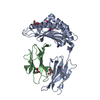
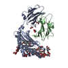
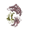
| Title | Structure of mouse CD1d in complex with PBS-25 | |||||||||
 Components Components |
| |||||||||
 Keywords Keywords | IMMUNE SYSTEM / CD1 / NKT cell / glycolipid / antigen presentation | |||||||||
| Function / homology |  Function and homology information Function and homology informationregulation of immature T cell proliferation in thymus / lipid antigen binding / positive regulation of NK T cell activation / positive regulation of NK T cell differentiation / NK T cell differentiation / endogenous lipid antigen binding / exogenous lipid antigen binding / antigen processing and presentation, endogenous lipid antigen via MHC class Ib / lipopeptide binding / antigen processing and presentation, exogenous lipid antigen via MHC class Ib ...regulation of immature T cell proliferation in thymus / lipid antigen binding / positive regulation of NK T cell activation / positive regulation of NK T cell differentiation / NK T cell differentiation / endogenous lipid antigen binding / exogenous lipid antigen binding / antigen processing and presentation, endogenous lipid antigen via MHC class Ib / lipopeptide binding / antigen processing and presentation, exogenous lipid antigen via MHC class Ib / positive thymic T cell selection / positive regulation of macrophage activation / Endosomal/Vacuolar pathway / DAP12 interactions / Antigen Presentation: Folding, assembly and peptide loading of class I MHC / DAP12 signaling / ER-Phagosome pathway / Immunoregulatory interactions between a Lymphoid and a non-Lymphoid cell / antigen processing and presentation / regulation of membrane depolarization / positive regulation of interleukin-4 production / regulation of immune response / positive regulation of interleukin-2 production / cellular defense response / T cell receptor binding / Neutrophil degranulation / positive regulation of T cell proliferation / cell adhesion molecule binding / regulation of iron ion transport / cellular response to iron(III) ion / negative regulation of iron ion transport / negative regulation of forebrain neuron differentiation / antigen processing and presentation of exogenous protein antigen via MHC class Ib, TAP-dependent / iron ion transport / peptide antigen assembly with MHC class I protein complex / regulation of erythrocyte differentiation / response to molecule of bacterial origin / HFE-transferrin receptor complex / MHC class I peptide loading complex / transferrin transport / cellular response to iron ion / negative regulation of receptor-mediated endocytosis / positive regulation of T cell cytokine production / antigen processing and presentation of endogenous peptide antigen via MHC class I / MHC class I protein complex / peptide antigen assembly with MHC class II protein complex / negative regulation of neurogenesis / cellular response to nicotine / MHC class II protein complex / positive regulation of receptor-mediated endocytosis / multicellular organismal-level iron ion homeostasis / positive regulation of T cell mediated cytotoxicity / antigen processing and presentation of exogenous peptide antigen via MHC class II / positive regulation of immune response / peptide antigen binding / positive regulation of type II interferon production / phagocytic vesicle membrane / positive regulation of T cell activation / negative regulation of epithelial cell proliferation / sensory perception of smell / positive regulation of cellular senescence / MHC class II protein complex binding / T cell differentiation in thymus / late endosome / late endosome membrane / antimicrobial humoral immune response mediated by antimicrobial peptide / negative regulation of neuron projection development / antibacterial humoral response / protein refolding / cellular response to lipopolysaccharide / amyloid fibril formation / protein homotetramerization / defense response to Gram-negative bacterium / intracellular iron ion homeostasis / learning or memory / early endosome / lysosome / endosome membrane / defense response to Gram-positive bacterium / immune response / external side of plasma membrane / innate immune response / lysosomal membrane / structural molecule activity / cell surface / endoplasmic reticulum / Golgi apparatus / protein homodimerization activity / : / identical protein binding / plasma membrane / cytoplasm / cytosol Similarity search - Function | |||||||||
| Biological species |  | |||||||||
| Method |  X-RAY DIFFRACTION / SYNCHROTRON / MOLECULAR REPLACEMENT / Resolution: 1.7 Å X-RAY DIFFRACTION / SYNCHROTRON / MOLECULAR REPLACEMENT / Resolution: 1.7 Å | |||||||||
 Authors Authors | Schiefner, A. / Wilson, I.A. | |||||||||
 Citation Citation | Journal: J.Mol.Biol. / Year: 2009 Title: Structural evaluation of potent NKT cell agonists: implications for design of novel stimulatory ligands. Authors: Schiefner, A. / Fujio, M. / Wu, D. / Wong, C.H. / Wilson, I.A. | |||||||||
| History |
|
- Structure visualization
| Structure viewer | Molecule: MolmilJmol/JSmol |
|---|
- Downloads & links
-Download
| PDBx/mmCIF format | 3gmp.cif.gz | 107.2 KB | Display | PDBx/mmCIF format |
|---|---|---|---|---|
| PDB format | pdb3gmp.ent.gz | 79.8 KB | Display | PDB format |
| PDBx/mmJSON format | 3gmp.json.gz | Tree view | PDBx/mmJSON format | |
| Others |  Other downloads Other downloads |
-Validation report
| Arichive directory | https://data.pdbj.org/pub/pdb/validation_reports/gm/3gmpftp://data.pdbj.org/pub/pdb/validation_reports/gm/3gmp | HTTPS FTP |
|---|
-Related structure data
| Related structure data |  3gmlSC  3gmmC  3gmnC  3gmoC  3gmqC  3gmrC C: citing same article ( S: Starting model for refinement |
|---|---|
| Similar structure data |


-Links
 PDBj
PDBj


- Assembly
| Deposited unit | 
| ||||||||
|---|---|---|---|---|---|---|---|---|---|
| 1 |
| ||||||||
| Unit cell |
|
-Components
-Protein , 2 types, 2 molecules AB
| #1: Protein | Mass: 32776.797 Da / Num. of mol.: 1 / Fragment: UNP residues 19-297 Source method: isolated from a genetically manipulated source Source: (gene. exp.)   Spodoptera frugiperda (fall armyworm) / Strain (production host): SF9 / References: UniProt: P11609 Spodoptera frugiperda (fall armyworm) / Strain (production host): SF9 / References: UniProt: P11609 |
|---|---|
| #2: Protein | Mass: 11660.350 Da / Num. of mol.: 1 / Fragment: UNP residues 21-119 Source method: isolated from a genetically manipulated source Source: (gene. exp.) Spodoptera frugiperda (fall armyworm) / Strain (production host): SF9 / References: UniProt: Q91XJ8, UniProt: P01887*PLUS |
-Sugars , 3 types, 3 molecules 
| #3: Polysaccharide | alpha-D-mannopyranose-(1-2)-alpha-D-mannopyranose-(1-3)-beta-D-mannopyranose-(1-4)-2-acetamido-2- ...alpha-D-mannopyranose-(1-2)-alpha-D-mannopyranose-(1-3)-beta-D-mannopyranose-(1-4)-2-acetamido-2-deoxy-beta-D-glucopyranose-(1-4)-2-acetamido-2-deoxy-beta-D-glucopyranose Source method: isolated from a genetically manipulated source |
|---|---|
| #4: Polysaccharide | alpha-D-mannopyranose-(1-3)-[alpha-D-mannopyranose-(1-6)]beta-D-mannopyranose-(1-4)-2-acetamido-2- ...alpha-D-mannopyranose-(1-3)-[alpha-D-mannopyranose-(1-6)]beta-D-mannopyranose-(1-4)-2-acetamido-2-deoxy-beta-D-glucopyranose-(1-4)-[alpha-L-fucopyranose-(1-6)]2-acetamido-2-deoxy-beta-D-glucopyranose Source method: isolated from a genetically manipulated source |
| #5: Sugar | ChemComp-NAG /  Type: D-saccharide, beta linking / Mass: 221.208 Da / Num. of mol.: 1 Type: D-saccharide, beta linking / Mass: 221.208 Da / Num. of mol.: 1Source method: isolated from a genetically manipulated source Formula: C8H15NO6 |
-Non-polymers , 4 types, 313 molecules 


| #6: Chemical | ChemComp-PBS / ( Mass: 605.844 Da / Num. of mol.: 1 / Source method: obtained synthetically / Formula: C32H63NO9 Mass: 605.844 Da / Num. of mol.: 1 / Source method: obtained synthetically / Formula: C32H63NO9 | ||
|---|---|---|---|
| #7: Chemical | ChemComp-PLM /  Mass: 256.424 Da / Num. of mol.: 1 / Source method: obtained synthetically / Formula: C16H32O2 Mass: 256.424 Da / Num. of mol.: 1 / Source method: obtained synthetically / Formula: C16H32O2 | ||
| #8: Chemical | ChemComp-EDO /  Mass: 62.068 Da / Num. of mol.: 4 / Source method: obtained synthetically / Formula: C2H6O2 Mass: 62.068 Da / Num. of mol.: 4 / Source method: obtained synthetically / Formula: C2H6O2#9: Water | ChemComp-HOH / | Mass: 18.015 Da / Num. of mol.: 307 / Source method: isolated from a natural source / Formula: H2O |
-Details
| Has protein modification | Y |
|---|---|
| Sequence details | ASP TO HIS CONFLICT IN UNP ENTRY P11609 |
-Experimental details
-Experiment
| Experiment | Method: X-RAY DIFFRACTION / Number of used crystals: 1 |
|---|
- Sample preparation
| Crystal | Density Matthews: 2.44 Å3/Da / Density % sol: 49.59 % |
|---|---|
| Crystal grow | Temperature: 295 K / Method: vapor diffusion, sitting drop / pH: 4.5 Details: 0.2 M Malonate pH 4.5, 20%(v/v) PEG3350, VAPOR DIFFUSION, SITTING DROP, temperature 295K |
-Data collection
| Diffraction | Mean temperature: 100 K |
|---|---|
| Diffraction source | Source: SYNCHROTRON / Site: SSRL  / Beamline: BL11-1 / Wavelength: 0.9795 Å / Beamline: BL11-1 / Wavelength: 0.9795 Å |
| Detector | Type: MARMOSAIC 325 mm CCD / Detector: CCD / Date: Apr 14, 2008 / Details: flat mirror |
| Radiation | Monochromator: Si(111) / Protocol: SINGLE WAVELENGTH / Monochromatic (M) / Laue (L): M / Scattering type: x-ray |
| Radiation wavelength | Wavelength: 0.9795 Å / Relative weight: 1 |
| Reflection | Resolution: 1.7→30 Å / Num. all: 46664 / Num. obs: 46664 / % possible obs: 99.5 % / Observed criterion σ(I): -3 / Redundancy: 4.1 % / Biso Wilson estimate: 31.9 Å2 / Rsym value: 0.031 / Net I/σ(I): 23.3 |
| Reflection shell | Resolution: 1.7→1.8 Å / Redundancy: 3.9 % / Mean I/σ(I) obs: 4 / Num. unique all: 7300 / Rsym value: 0.336 / % possible all: 98.8 |
- Processing
| Software |
| ||||||||||||||||||||||||||||||||||||||||||||||||||||||||||||||||||||||||||||||||||||||||||||||||||||
|---|---|---|---|---|---|---|---|---|---|---|---|---|---|---|---|---|---|---|---|---|---|---|---|---|---|---|---|---|---|---|---|---|---|---|---|---|---|---|---|---|---|---|---|---|---|---|---|---|---|---|---|---|---|---|---|---|---|---|---|---|---|---|---|---|---|---|---|---|---|---|---|---|---|---|---|---|---|---|---|---|---|---|---|---|---|---|---|---|---|---|---|---|---|---|---|---|---|---|---|---|---|
| Refinement | Method to determine structure: MOLECULAR REPLACEMENT Starting model: PDB entry 3GML Resolution: 1.7→27.78 Å / Cor.coef. Fo:Fc: 0.962 / Cor.coef. Fo:Fc free: 0.946 / SU B: 4.442 / SU ML: 0.07 / TLS residual ADP flag: LIKELY RESIDUAL / Cross valid method: THROUGHOUT / σ(F): 0 / ESU R: 0.105 / ESU R Free: 0.106 / Stereochemistry target values: MAXIMUM LIKELIHOOD / Details: HYDROGENS HAVE BEEN ADDED IN THE RIDING POSITIONS
| ||||||||||||||||||||||||||||||||||||||||||||||||||||||||||||||||||||||||||||||||||||||||||||||||||||
| Solvent computation | Ion probe radii: 0.8 Å / Shrinkage radii: 0.8 Å / VDW probe radii: 1.4 Å / Solvent model: MASK | ||||||||||||||||||||||||||||||||||||||||||||||||||||||||||||||||||||||||||||||||||||||||||||||||||||
| Displacement parameters | Biso mean: 23.566 Å2
| ||||||||||||||||||||||||||||||||||||||||||||||||||||||||||||||||||||||||||||||||||||||||||||||||||||
| Refinement step | Cycle: LAST / Resolution: 1.7→27.78 Å
| ||||||||||||||||||||||||||||||||||||||||||||||||||||||||||||||||||||||||||||||||||||||||||||||||||||
| Refine LS restraints |
| ||||||||||||||||||||||||||||||||||||||||||||||||||||||||||||||||||||||||||||||||||||||||||||||||||||
| LS refinement shell | Resolution: 1.7→1.744 Å / Total num. of bins used: 20
| ||||||||||||||||||||||||||||||||||||||||||||||||||||||||||||||||||||||||||||||||||||||||||||||||||||
| Refinement TLS params. | Method: refined / Refine-ID: X-RAY DIFFRACTION
| ||||||||||||||||||||||||||||||||||||||||||||||||||||||||||||||||||||||||||||||||||||||||||||||||||||
| Refinement TLS group |
|
