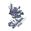
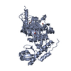
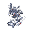
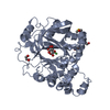
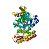
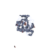
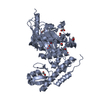
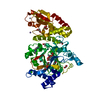
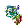
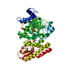
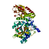
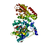
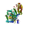
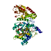
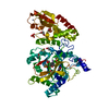
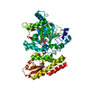
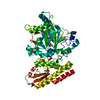
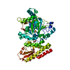
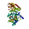
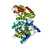
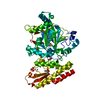
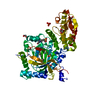
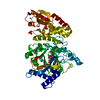
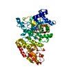
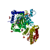
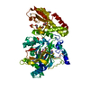
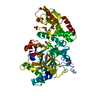
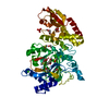
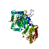
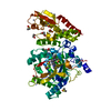
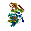
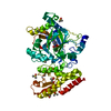
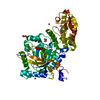
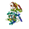
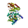
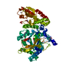
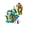
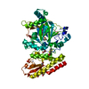
- PDB-5fz4: Crystal structure of the catalytic domain of human JARID1B in com... -
+
Open data
ID or keywords:
Loading...
-
Basic information
Entry
Database: PDB / ID: 5fz4
Title
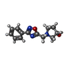
Crystal structure of the catalytic domain of human JARID1B in complex with 3D fragment (3R)-1-[(3-phenyl-1,2,4-oxadiazol-5-yl)methyl]pyrrolidin-3-ol (N10057a) (ligand modelled based on PANDDA event map, SGC - Diamond I04-1 fragment screening)
Mass: 55214.297 Da / Num. of mol.: 1 / Fragment: JMJC DOMAIN, RESIDUES 26-101,374-770 Source method: isolated from a genetically manipulated source Source: (gene. exp.) HOMO SAPIENS (human) / Plasmid: PFB-LIC-BSE / Cell line (production host): SF9 / Production host: SPODOPTERA FRUGIPERDA (fall armyworm) References: UniProt: Q9UGL1, Oxidoreductases; Acting on paired donors, with incorporation or reduction of molecular oxygen; With 2-oxoglutarate as one donor, and incorporation of one atom of oxygen into each donor
Mass: 18.015 Da / Num. of mol.: 205 / Source method: isolated from a natural source / Formula: H2O
-
Details
Sequence details
RESIDUES S AND M COME FROM THE EXPRESSION VECTOR. RESIDUES 102-373 WERE DELETED FROM THE ORIGINAL ...RESIDUES S AND M COME FROM THE EXPRESSION VECTOR. RESIDUES 102-373 WERE DELETED FROM THE ORIGINAL SEQUENCE Q9UGL1. DELETED RESIDUES WERE LINKED WITH 4 GLYCINE LINKER (GGGG).
-
Experimental details
-
Experiment
Experiment
Method: X-RAY DIFFRACTION / Number of used crystals: 1
-
Sample preparation
Crystal
Density Matthews: 2.1 Å3/Da / Density % sol: 41.4 % / Description: NONE
Resolution: 2.07→70.68 Å / SU ML: 0.27 / σ(F): 1.34 / Phase error: 25.2 / Stereochemistry target values: ML Details: LIGAND MODELLED BASED ON PANDDA EVENT MAP TO BE PUBLISHED.
Rfactor
Num. reflection
% reflection
Rfree
0.2343
2672
4.9 %
Rwork
0.1984
-
-
obs
0.2002
54952
99.99 %
Solvent computation
Shrinkage radii: 0.9 Å / VDW probe radii: 1.11 Å / Solvent model: FLAT BULK SOLVENT MODEL
Refinement step
Cycle: LAST / Resolution: 2.07→70.68 Å
Protein
Nucleic acid
Ligand
Solvent
Total
Num. atoms
3651
0
79
205
3935
Refine LS restraints
Refine-ID
Type
Dev ideal
Number
X-RAY DIFFRACTION
f_bond_d
0.009
3920
X-RAY DIFFRACTION
f_angle_d
1.196
5315
X-RAY DIFFRACTION
f_dihedral_angle_d
15.121
1474
X-RAY DIFFRACTION
f_chiral_restr
0.048
558
X-RAY DIFFRACTION
f_plane_restr
0.006
683
LS refinement shell
Resolution (Å)
Rfactor Rfree
Num. reflection Rfree
Rfactor Rwork
Num. reflection Rwork
Refine-ID
% reflection obs (%)
2.07-2.1077
0.3442
145
0.3223
2686
X-RAY DIFFRACTION
100
2.1077-2.1482
0.2987
134
0.3074
2717
X-RAY DIFFRACTION
100
2.1482-2.192
0.2982
131
0.2753
2696
X-RAY DIFFRACTION
100
2.192-2.2397
0.3492
132
0.2781
2719
X-RAY DIFFRACTION
100
2.2397-2.2918
0.3117
135
0.256
2707
X-RAY DIFFRACTION
100
2.2918-2.3491
0.2821
129
0.2441
2720
X-RAY DIFFRACTION
100
2.3491-2.4127
0.2889
146
0.243
2721
X-RAY DIFFRACTION
100
2.4127-2.4836
0.2615
146
0.2418
2713
X-RAY DIFFRACTION
100
2.4836-2.5638
0.2889
117
0.2314
2742
X-RAY DIFFRACTION
100
2.5638-2.6555
0.2791
149
0.2227
2710
X-RAY DIFFRACTION
100
2.6555-2.7618
0.272
144
0.2219
2723
X-RAY DIFFRACTION
100
2.7618-2.8875
0.2568
155
0.2134
2729
X-RAY DIFFRACTION
100
2.8875-3.0397
0.2597
125
0.2131
2755
X-RAY DIFFRACTION
100
3.0397-3.2302
0.2097
148
0.2112
2751
X-RAY DIFFRACTION
100
3.2302-3.4796
0.2572
106
0.201
2787
X-RAY DIFFRACTION
100
3.4796-3.8297
0.2163
159
0.1905
2779
X-RAY DIFFRACTION
100
3.8297-4.3838
0.2085
155
0.1662
2780
X-RAY DIFFRACTION
100
4.3838-5.5228
0.1898
144
0.1509
2842
X-RAY DIFFRACTION
100
5.5228-70.7212
0.2171
172
0.1802
3003
X-RAY DIFFRACTION
100
+
About Yorodumi
-
News
-
Feb 9, 2022. New format data for meta-information of EMDB entries
New format data for meta-information of EMDB entries
Version 3 of the EMDB header file is now the official format.
The previous official version 1.9 will be removed from the archive.
In the structure databanks used in Yorodumi, some data are registered as the other names, "COVID-19 virus" and "2019-nCoV". Here are the details of the virus and the list of structure data.
Jan 31, 2019. EMDB accession codes are about to change! (news from PDBe EMDB page)
EMDB accession codes are about to change! (news from PDBe EMDB page)
The allocation of 4 digits for EMDB accession codes will soon come to an end. Whilst these codes will remain in use, new EMDB accession codes will include an additional digit and will expand incrementally as the available range of codes is exhausted. The current 4-digit format prefixed with “EMD-” (i.e. EMD-XXXX) will advance to a 5-digit format (i.e. EMD-XXXXX), and so on. It is currently estimated that the 4-digit codes will be depleted around Spring 2019, at which point the 5-digit format will come into force.
The EM Navigator/Yorodumi systems omit the EMD- prefix.
Related info.:Q: What is EMD? / ID/Accession-code notation in Yorodumi/EM Navigator
Yorodumi is a browser for structure data from EMDB, PDB, SASBDB, etc.
This page is also the successor to EM Navigator detail page, and also detail information page/front-end page for Omokage search.
The word "yorodu" (or yorozu) is an old Japanese word meaning "ten thousand". "mi" (miru) is to see.
Related info.:EMDB / PDB / SASBDB / Comparison of 3 databanks / Yorodumi Search / Aug 31, 2016. New EM Navigator & Yorodumi / Yorodumi Papers / Jmol/JSmol / Function and homology information / Changes in new EM Navigator and Yorodumi
 Movie
Movie Controller

 Yorodumi
 Open data
 Basic information
 Components
 Keywords
 Function and homology information
 HOMO SAPIENS (human)
 X-RAY DIFFRACTION /
  Authors
 Citation
 Structure visualization
 Downloads & links
 Other downloads
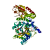


 PDBj
PDBj

 Assembly
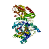

 SPODOPTERA FRUGIPERDA (fall armyworm)


 Mass: 65.409 Da / Num. of mol.: 2 / Source method: obtained synthetically / Formula: Zn
 Mass: 245.277 Da / Num. of mol.: 1 / Source method: obtained synthetically / Formula: C13H15N3O2
 Mass: 54.938 Da / Num. of mol.: 1 / Source method: obtained synthetically / Formula: Mn
 Mass: 35.453 Da / Num. of mol.: 1 / Source method: obtained synthetically / Formula: Cl
 Mass: 62.068 Da / Num. of mol.: 10 / Source method: obtained synthetically / Formula: C2H6O2
 Mass: 78.133 Da / Num. of mol.: 3 / Source method: obtained synthetically / Formula: C2H6OS / Comment: DMSO, precipitant*YM
 Mass: 94.971 Da / Num. of mol.: 1 / Source method: obtained synthetically / Formula: PO4
 Sample preparation
 / Beamline: I04-1 / Wavelength: 0.91732
  Processing