 Movie
Movie Controller

[English] 日本語
 Yorodumi
- PDB-5fv3: Crystal structure of human JARID1B construct c2 in complex with N... -
+ Open data

- Basic information
| Entry | Database: PDB / ID: 5fv3 | ||||||
|---|---|---|---|---|---|---|---|
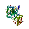
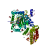
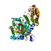
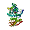
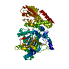
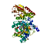
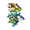
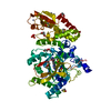
| Title | Crystal structure of human JARID1B construct c2 in complex with N- Oxalylglycine. | ||||||
 Components Components | LYSINE-SPECIFIC DEMETHYLASE 5B, LYSINE-SPECIFIC DEMETHYLASE 5B | ||||||
 Keywords Keywords | OXIDOREDUCTASE / JARID1B / PLU1 | ||||||
| Function / homology |  Function and homology information Function and homology informationregulation of estradiol secretion / mammary duct terminal end bud growth / uterus morphogenesis / TFAP2 (AP-2) family regulates transcription of cell cycle factors / positive regulation of mammary gland epithelial cell proliferation / [histone H3]-trimethyl-L-lysine4 demethylase / histone H3K4me/H3K4me2/H3K4me3 demethylase activity / lens fiber cell differentiation / histone H3K4 demethylase activity / branching involved in mammary gland duct morphogenesis ...regulation of estradiol secretion / mammary duct terminal end bud growth / uterus morphogenesis / TFAP2 (AP-2) family regulates transcription of cell cycle factors / positive regulation of mammary gland epithelial cell proliferation / [histone H3]-trimethyl-L-lysine4 demethylase / histone H3K4me/H3K4me2/H3K4me3 demethylase activity / lens fiber cell differentiation / histone H3K4 demethylase activity / branching involved in mammary gland duct morphogenesis / histone demethylase activity / single fertilization / response to fungicide / cellular response to fibroblast growth factor stimulus / cellular response to leukemia inhibitory factor / Chromatin modifications during the maternal to zygotic transition (MZT) / post-embryonic development / HDMs demethylate histones / sequence-specific double-stranded DNA binding / transcription corepressor activity / rhythmic process / histone binding / nucleic acid binding / chromatin remodeling / negative regulation of DNA-templated transcription / positive regulation of gene expression / regulation of DNA-templated transcription / chromatin / DNA binding / zinc ion binding / nucleoplasm / nucleus / cytosol Similarity search - Function | ||||||
| Biological species |  HOMO SAPIENS (human) HOMO SAPIENS (human) | ||||||
| Method |  X-RAY DIFFRACTION / SYNCHROTRON / MOLECULAR REPLACEMENT / Resolution: 2.37 Å X-RAY DIFFRACTION / SYNCHROTRON / MOLECULAR REPLACEMENT / Resolution: 2.37 Å | ||||||
 Authors Authors | Nowak, R. / Srikannathasan, V. / Johansson, C. / Gileadi, C. / Kupinska, K. / Strain-Damerell, C. / Szykowska, A. / Talon, R. / von Delft, F. / Burgess-Brown, N.A. ...Nowak, R. / Srikannathasan, V. / Johansson, C. / Gileadi, C. / Kupinska, K. / Strain-Damerell, C. / Szykowska, A. / Talon, R. / von Delft, F. / Burgess-Brown, N.A. / Arrowsmith, C.H. / Bountra, C. / Edwards, A.M. / Oppermann, U. | ||||||
 Citation Citation | Journal: Nat.Chem.Biol. / Year: 2016 Title: Structural Analysis of Human Kdm5B Guides Histone Demethylase Inhibitor Development. Authors: Johansson, C. / Velupillai, S. / Tumber, A. / Szykowska, A. / Hookway, E.S. / Nowak, R.P. / Strain-Damerell, C. / Gileadi, C. / Philpott, M. / Burgess-Brown, N. / Wu, N. / Kopec, J. / Nuzzi, ...Authors: Johansson, C. / Velupillai, S. / Tumber, A. / Szykowska, A. / Hookway, E.S. / Nowak, R.P. / Strain-Damerell, C. / Gileadi, C. / Philpott, M. / Burgess-Brown, N. / Wu, N. / Kopec, J. / Nuzzi, A. / Steuber, H. / Egner, U. / Badock, V. / Munro, S. / Lathangue, N.B. / Westaway, S. / Brown, J. / Athanasou, N. / Prinjha, R. / Brennan, P.E. / Oppermann, U. | ||||||
| History |
|
- Structure visualization
| Structure viewer | Molecule: MolmilJmol/JSmol |
|---|
- Downloads & links
-Download
| PDBx/mmCIF format | 5fv3.cif.gz | 115.6 KB | Display | PDBx/mmCIF format |
|---|---|---|---|---|
| PDB format | pdb5fv3.ent.gz | 87.4 KB | Display | PDB format |
| PDBx/mmJSON format | 5fv3.json.gz | Tree view | PDBx/mmJSON format | |
| Others |  Other downloads Other downloads |
-Validation report
| Arichive directory | https://data.pdbj.org/pub/pdb/validation_reports/fv/5fv3ftp://data.pdbj.org/pub/pdb/validation_reports/fv/5fv3 | HTTPS FTP |
|---|
-Related structure data
| Related structure data |  4uf0C  5a1fSC  5a3pC  5a3tC  5a3wC  5fpuC  5fpvC  5funC  5fupC  5fwjC C: citing same article ( S: Starting model for refinement |
|---|---|
| Similar structure data |


-Links
 PDBj
PDBj

- Assembly
| Deposited unit | 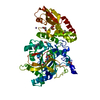
| ||||||||
|---|---|---|---|---|---|---|---|---|---|
| 1 |
| ||||||||
| Unit cell |
| ||||||||
| Components on special symmetry positions |
|
-Components
-Protein , 1 types, 1 molecules A
| #1: Protein | Mass: 55746.844 Da / Num. of mol.: 1 Fragment: JMJC DOMAIN, RESIDUES 26-101, JMJC DOMAIN, RESIDUES 374-770 Source method: isolated from a genetically manipulated source Source: (gene. exp.) HOMO SAPIENS (human) / Plasmid: PFB-LIC-BSE / Cell line (production host): SF9 / Production host:   SPODOPTERA FRUGIPERDA (fall armyworm) SPODOPTERA FRUGIPERDA (fall armyworm)References: UniProt: Q9UGL1, Oxidoreductases; Acting on paired donors, with incorporation or reduction of molecular oxygen; With 2-oxoglutarate as one donor, and incorporation of one atom of oxygen into each donor |
|---|
-Non-polymers , 8 types, 137 molecules 


| #2: Chemical | ChemComp-ZN /  Mass: 65.409 Da / Num. of mol.: 1 / Source method: obtained synthetically / Formula: Zn Mass: 65.409 Da / Num. of mol.: 1 / Source method: obtained synthetically / Formula: Zn | ||||||||||||
|---|---|---|---|---|---|---|---|---|---|---|---|---|---|
| #3: Chemical |  Mass: 78.133 Da / Num. of mol.: 2 / Source method: obtained synthetically / Formula: C2H6OS / Comment: DMSO, precipitant*YM Mass: 78.133 Da / Num. of mol.: 2 / Source method: obtained synthetically / Formula: C2H6OS / Comment: DMSO, precipitant*YM#4: Chemical |  Mass: 54.938 Da / Num. of mol.: 2 / Source method: obtained synthetically / Formula: Mn Mass: 54.938 Da / Num. of mol.: 2 / Source method: obtained synthetically / Formula: Mn#5: Chemical | ChemComp-EPE / |  Mass: 238.305 Da / Num. of mol.: 1 / Source method: obtained synthetically / Formula: C8H18N2O4S / Comment: pH buffer*YM Mass: 238.305 Da / Num. of mol.: 1 / Source method: obtained synthetically / Formula: C8H18N2O4S / Comment: pH buffer*YM#6: Chemical | ChemComp-EDO /  Mass: 62.068 Da / Num. of mol.: 9 / Source method: obtained synthetically / Formula: C2H6O2 Mass: 62.068 Da / Num. of mol.: 9 / Source method: obtained synthetically / Formula: C2H6O2#7: Chemical | ChemComp-CL / |  Mass: 35.453 Da / Num. of mol.: 1 / Source method: obtained synthetically / Formula: Cl Mass: 35.453 Da / Num. of mol.: 1 / Source method: obtained synthetically / Formula: Cl#8: Chemical | ChemComp-OGA / |  Mass: 147.086 Da / Num. of mol.: 1 / Source method: obtained synthetically / Formula: C4H5NO5 / Comment: inhibitor*YM Mass: 147.086 Da / Num. of mol.: 1 / Source method: obtained synthetically / Formula: C4H5NO5 / Comment: inhibitor*YM#9: Water | ChemComp-HOH / | Mass: 18.015 Da / Num. of mol.: 120 / Source method: isolated from a natural source / Formula: H2O |
-Details
| Has protein modification | Y |
|---|---|
| Sequence details | RESIDUE S AND M FROM THE EXPRESSION PLASMID, SEQUENCE N102 (INCLUDING) TO F368 (INCLUDING) WAS ...RESIDUE S AND M FROM THE EXPRESSION |
-Experimental details
-Experiment
| Experiment | Method: X-RAY DIFFRACTION / Number of used crystals: 1 |
|---|
- Sample preparation
| Crystal | Density Matthews: 2 Å3/Da / Density % sol: 38.66 % / Description: NONE |
|---|---|
| Crystal grow | pH: 7.5 Details: 0.1M HEPES PH 7.5, 0.8M POTASSIUM PHOSPHATE DIBASIC, 0.8M SODIUM PHOSPHATE MONOBASIC |
-Data collection
| Diffraction | Mean temperature: 100 K |
|---|---|
| Diffraction source | Source: SYNCHROTRON / Site: Diamond  / Beamline: I03 / Wavelength: 0.97626 / Beamline: I03 / Wavelength: 0.97626 |
| Detector | Type: DECTRIS PILATUS 6M / Detector: PIXEL / Date: Jul 2, 2015 |
| Radiation | Protocol: SINGLE WAVELENGTH / Monochromatic (M) / Laue (L): M / Scattering type: x-ray |
| Radiation wavelength | Wavelength: 0.97626 Å / Relative weight: 1 |
| Reflection | Resolution: 2.37→123.28 Å / Num. obs: 37525 / % possible obs: 100 % / Observed criterion σ(I): 1.5 / Redundancy: 14.1 % / Biso Wilson estimate: 47.84 Å2 / Rmerge(I) obs: 0.31 / Net I/σ(I): 8.8 |
| Reflection shell | Resolution: 2.37→2.43 Å / Redundancy: 15.2 % / Rmerge(I) obs: 1.5 / Mean I/σ(I) obs: 1.2 / % possible all: 100 |
- Processing
| Software |
| ||||||||||||||||||||||||||||||||||||||||||||||||||||||||||||||||||||||||||||||||||||||||||||||||||
|---|---|---|---|---|---|---|---|---|---|---|---|---|---|---|---|---|---|---|---|---|---|---|---|---|---|---|---|---|---|---|---|---|---|---|---|---|---|---|---|---|---|---|---|---|---|---|---|---|---|---|---|---|---|---|---|---|---|---|---|---|---|---|---|---|---|---|---|---|---|---|---|---|---|---|---|---|---|---|---|---|---|---|---|---|---|---|---|---|---|---|---|---|---|---|---|---|---|---|---|
| Refinement | Method to determine structure: MOLECULAR REPLACEMENT Starting model: PDB ENTRY 5A1F Resolution: 2.37→123.279 Å / SU ML: 0.36 / σ(F): 1.34 / Phase error: 26.74 / Stereochemistry target values: ML Details: SIDE CHAINS WITHOUT DENSITY WERE REMOVED. SOME RESIDUES HAVE ALTERNATIVE CONFORMATION. GLYCINE LINKER DOES NOT EXIST.
| ||||||||||||||||||||||||||||||||||||||||||||||||||||||||||||||||||||||||||||||||||||||||||||||||||
| Solvent computation | Shrinkage radii: 0.9 Å / VDW probe radii: 1.11 Å / Solvent model: FLAT BULK SOLVENT MODEL / Bsol: 2 Å2 / ksol: 2 e/Å3 | ||||||||||||||||||||||||||||||||||||||||||||||||||||||||||||||||||||||||||||||||||||||||||||||||||
| Refinement step | Cycle: LAST / Resolution: 2.37→123.279 Å
| ||||||||||||||||||||||||||||||||||||||||||||||||||||||||||||||||||||||||||||||||||||||||||||||||||
| Refine LS restraints |
| ||||||||||||||||||||||||||||||||||||||||||||||||||||||||||||||||||||||||||||||||||||||||||||||||||
| LS refinement shell |
|
