Resolution: 1.8→40 Å / Cor.coef. Fo:Fc: 0.969 / SU B: 1.788 / SU ML: 0.055 / Isotropic thermal model: isotropic / Cross valid method: Rfree throughout / σ(F): 0 Stereochemistry target values: CCP4 6.1.3 stereochemistry library Details: HYDROGENS HAVE BEEN GENERATED AND USED ONLY IN REFINEMENT. STRUCTURE SOLUTION AND REFINEMENT WERE PERFORMED USING FREE R FOR CROSS-VALIDATION THROUGHOUT: FREE R = 0.185 (FREE R = 0.2610 FOR ...Details: HYDROGENS HAVE BEEN GENERATED AND USED ONLY IN REFINEMENT. STRUCTURE SOLUTION AND REFINEMENT WERE PERFORMED USING FREE R FOR CROSS-VALIDATION THROUGHOUT: FREE R = 0.185 (FREE R = 0.2610 FOR THE HIGHEST RESOLUTION SHELL) FROM A RANDOM TEST SET COMPRISING 5% OF REFLECTIONS (47975 TOTAL). FINAL REFINEMENT WAS PERFORMED USING ALL REFLECTIONS.
Rfactor
Num. reflection
% reflection
Selection details
Rwork
0.1559
-
-
-
all
0.1563
47975
-
-
obs
0.1563
47975
99.43 %
-
Rfree
-
2371
-
random
Solvent computation
Ion probe radii: 0.8 Å / Shrinkage radii: 0.8 Å / VDW probe radii: 1.2 Å / Solvent model: MASK
Displacement parameters
Biso mean: 18.099 Å2
Baniso -1
Baniso -2
Baniso -3
1-
-0.88 Å2
0 Å2
-1.39 Å2
2-
-
0.05 Å2
0 Å2
3-
-
-
-0.76 Å2
Refinement step
Cycle: LAST / Resolution: 1.8→40 Å
Protein
Nucleic acid
Ligand
Solvent
Total
Num. atoms
3579
0
8
464
4051
Refine LS restraints
Refine-ID
Type
Dev ideal
Dev ideal target
Number
X-RAY DIFFRACTION
r_bond_refined_d
0.014
0.022
3776
X-RAY DIFFRACTION
r_bond_other_d
0.001
0.02
2543
X-RAY DIFFRACTION
r_angle_refined_deg
1.337
1.945
5151
X-RAY DIFFRACTION
r_angle_other_deg
0.87
3
6204
X-RAY DIFFRACTION
r_dihedral_angle_1_deg
9.504
5.043
466
X-RAY DIFFRACTION
r_dihedral_angle_2_deg
32.397
24.279
201
X-RAY DIFFRACTION
r_dihedral_angle_3_deg
12.064
15
627
X-RAY DIFFRACTION
r_dihedral_angle_4_deg
15.682
15
24
X-RAY DIFFRACTION
r_chiral_restr
0.087
0.2
558
X-RAY DIFFRACTION
r_gen_planes_refined
0.007
0.021
4249
X-RAY DIFFRACTION
r_gen_planes_other
0.001
0.02
778
LS refinement shell
Resolution: 1.802→1.849 Å / Total num. of bins used: 20
Rfactor
Num. reflection
% reflection
Rwork
0.24
3274
-
Rfree
-
159
-
obs
-
3274
95.34 %
+
About Yorodumi
-
News
-
Feb 9, 2022. New format data for meta-information of EMDB entries
New format data for meta-information of EMDB entries
Version 3 of the EMDB header file is now the official format.
The previous official version 1.9 will be removed from the archive.
In the structure databanks used in Yorodumi, some data are registered as the other names, "COVID-19 virus" and "2019-nCoV". Here are the details of the virus and the list of structure data.
Jan 31, 2019. EMDB accession codes are about to change! (news from PDBe EMDB page)
EMDB accession codes are about to change! (news from PDBe EMDB page)
The allocation of 4 digits for EMDB accession codes will soon come to an end. Whilst these codes will remain in use, new EMDB accession codes will include an additional digit and will expand incrementally as the available range of codes is exhausted. The current 4-digit format prefixed with “EMD-” (i.e. EMD-XXXX) will advance to a 5-digit format (i.e. EMD-XXXXX), and so on. It is currently estimated that the 4-digit codes will be depleted around Spring 2019, at which point the 5-digit format will come into force.
The EM Navigator/Yorodumi systems omit the EMD- prefix.
Related info.:Q: What is EMD? / ID/Accession-code notation in Yorodumi/EM Navigator
Yorodumi is a browser for structure data from EMDB, PDB, SASBDB, etc.
This page is also the successor to EM Navigator detail page, and also detail information page/front-end page for Omokage search.
The word "yorodu" (or yorozu) is an old Japanese word meaning "ten thousand". "mi" (miru) is to see.
Related info.:EMDB / PDB / SASBDB / Comparison of 3 databanks / Yorodumi Search / Aug 31, 2016. New EM Navigator & Yorodumi / Yorodumi Papers / Jmol/JSmol / Function and homology information / Changes in new EM Navigator and Yorodumi
 Movie
Movie Controller

 Yorodumi
 Open data
 Basic information
 Components
 Keywords
 Function and homology information
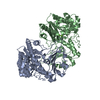
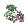
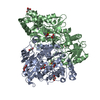
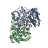
 Alteromonas macleodii (bacteria)
 X-RAY DIFFRACTION /
  Authors
 Citation
 Structure visualization
 Downloads & links
 Other downloads


 PDBj
PDBj Assembly
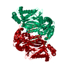
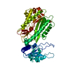

 Mass: 54.938 Da / Num. of mol.: 2 / Source method: obtained synthetically / Formula: Mn
 Mass: 94.971 Da / Num. of mol.: 1 / Source method: obtained synthetically / Formula: PO4
 Mass: 58.693 Da / Num. of mol.: 1 / Source method: obtained synthetically / Formula: Ni
 Mass: 18.015 Da / Num. of mol.: 464 / Source method: isolated from a natural source / Formula: H2O
 Sample preparation
 / Beamline: 14.1 / Wavelength: 0.91841 Å
 Processing