 Movie
Movie Controller

[English] 日本語
 Yorodumi
- PDB-1id1: CRYSTAL STRUCTURE OF THE RCK DOMAIN FROM E.COLI POTASSIUM CHANNEL -
+ Open data

- Basic information
| Entry | Database: PDB / ID: 1id1 | ||||||
|---|---|---|---|---|---|---|---|
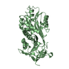
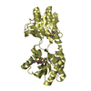
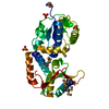
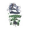
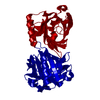
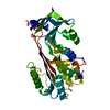
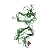
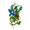
| Title | CRYSTAL STRUCTURE OF THE RCK DOMAIN FROM E.COLI POTASSIUM CHANNEL | ||||||
 Components Components | PUTATIVE POTASSIUM CHANNEL PROTEIN | ||||||
 Keywords Keywords | MEMBRANE PROTEIN / RCK domain / E.coli Potassium Channel / BK Channel / Rossmann fold | ||||||
| Function / homology |  Function and homology information Function and homology informationpotassium channel activity / potassium ion transmembrane transport / potassium ion transport / protein-containing complex / identical protein binding / plasma membrane Similarity search - Function | ||||||
| Biological species |  | ||||||
| Method |  X-RAY DIFFRACTION / SYNCHROTRON / MOLECULAR REPLACEMENT / Resolution: 2.4 Å X-RAY DIFFRACTION / SYNCHROTRON / MOLECULAR REPLACEMENT / Resolution: 2.4 Å | ||||||
 Authors Authors | Jiang, Y. / Pico, A. / Cadene, M. / Chait, B.T. / MacKinnon, R. | ||||||
 Citation Citation | Journal: Neuron / Year: 2001 Title: Structure of the RCK domain from the E. coli K+ channel and demonstration of its presence in the human BK channel. Authors: Jiang, Y. / Pico, A. / Cadene, M. / Chait, B.T. / MacKinnon, R. | ||||||
| History |
|
- Structure visualization
| Structure viewer | Molecule: MolmilJmol/JSmol |
|---|
- Downloads & links
-Download
| PDBx/mmCIF format | 1id1.cif.gz | 66.9 KB | Display | PDBx/mmCIF format |
|---|---|---|---|---|
| PDB format | pdb1id1.ent.gz | 50.7 KB | Display | PDB format |
| PDBx/mmJSON format | 1id1.json.gz | Tree view | PDBx/mmJSON format | |
| Others |  Other downloads Other downloads |
-Validation report
| Arichive directory | https://data.pdbj.org/pub/pdb/validation_reports/id/1id1ftp://data.pdbj.org/pub/pdb/validation_reports/id/1id1 | HTTPS FTP |
|---|
-Related structure data
| Similar structure data |
|---|


-Links
 PDBj
PDBj

- Assembly
| Deposited unit | 
| ||||||||
|---|---|---|---|---|---|---|---|---|---|
| 1 |
| ||||||||
| Unit cell |
|
-Components
| #1: Protein | Mass: 16746.068 Da / Num. of mol.: 2 / Fragment: C-TERMINAL DOMAIN, RESIDUES 241-393 Source method: isolated from a genetically manipulated source Source: (gene. exp.) #2: Water | ChemComp-HOH / |  Mass: 18.015 Da / Num. of mol.: 57 / Source method: isolated from a natural source / Formula: H2O Mass: 18.015 Da / Num. of mol.: 57 / Source method: isolated from a natural source / Formula: H2O |
|---|
-Experimental details
-Experiment
| Experiment | Method: X-RAY DIFFRACTION / Number of used crystals: 1 |
|---|
- Sample preparation
| Crystal | Density Matthews: 3.53 Å3/Da / Density % sol: 65.16 % | ||||||||||||||||||||||||||||||||||||||||||
|---|---|---|---|---|---|---|---|---|---|---|---|---|---|---|---|---|---|---|---|---|---|---|---|---|---|---|---|---|---|---|---|---|---|---|---|---|---|---|---|---|---|---|---|
| Crystal grow | Temperature: 293 K / Method: vapor diffusion, sitting drop / pH: 8 Details: PEG 4000, sodium chloride, tris, pH 8.0, VAPOR DIFFUSION, SITTING DROP, temperature 293K | ||||||||||||||||||||||||||||||||||||||||||
| Crystal grow | *PLUS Temperature: 20 ℃ | ||||||||||||||||||||||||||||||||||||||||||
| Components of the solutions | *PLUS
|
-Data collection
| Diffraction | Mean temperature: 100 K |
|---|---|
| Diffraction source | Source: SYNCHROTRON / Site: NSLS  / Beamline: X25 / Wavelength: 1.1 Å / Beamline: X25 / Wavelength: 1.1 Å |
| Detector | Type: BRANDEIS / Detector: CCD / Date: May 1, 1999 |
| Radiation | Protocol: SINGLE WAVELENGTH / Monochromatic (M) / Laue (L): M / Scattering type: x-ray |
| Radiation wavelength | Wavelength: 1.1 Å / Relative weight: 1 |
| Reflection shell | Resolution: 2.4→30 Å / Redundancy: 4.8 % / Rmerge(I) obs: 0.086 / % possible all: 99.5 |
| Reflection | *PLUS Highest resolution: 2.4 Å / Lowest resolution: 30 Å / % possible obs: 99.5 % / Redundancy: 4.8 % / Rmerge(I) obs: 0.086 |
| Reflection shell | *PLUS % possible obs: 96.3 % / Rmerge(I) obs: 0.292 |
- Processing
| Software |
| ||||||||||||||||||||
|---|---|---|---|---|---|---|---|---|---|---|---|---|---|---|---|---|---|---|---|---|---|
| Refinement | Method to determine structure: MOLECULAR REPLACEMENT / Resolution: 2.4→30 Å / Cross valid method: THROUGHOUT / σ(F): 0 / σ(I): 0 / Stereochemistry target values: Engh & Huber
| ||||||||||||||||||||
| Refinement step | Cycle: LAST / Resolution: 2.4→30 Å
| ||||||||||||||||||||
| Refine LS restraints |
|
