 Movie
Movie Controller

[English] 日本語
 Yorodumi
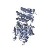
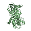
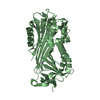
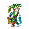
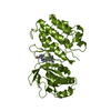
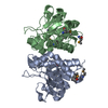
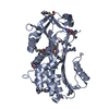
- PDB-1by7: HUMAN PLASMINOGEN ACTIVATOR INHIBITOR-2. LOOP (66-98) DELETION MUTANT -
+ Open data

- Basic information
| Entry | Database: PDB / ID: 1by7 | ||||||
|---|---|---|---|---|---|---|---|
| Title | HUMAN PLASMINOGEN ACTIVATOR INHIBITOR-2. LOOP (66-98) DELETION MUTANT | ||||||
 Components Components | PROTEIN (PLASMINOGEN ACTIVATOR INHIBITOR-2) | ||||||
 Keywords Keywords | PROTEIN BINDING / SERPIN | ||||||
| Function / homology |  Function and homology information Function and homology informationcornified envelope / Dissolution of Fibrin Clot / fibrinolysis / Gene and protein expression by JAK-STAT signaling after Interleukin-12 stimulation / serine-type endopeptidase inhibitor activity / negative regulation of apoptotic process / : / extracellular region / plasma membrane / cytoplasm Similarity search - Function | ||||||
| Biological species |  Homo sapiens (human) Homo sapiens (human) | ||||||
| Method |  X-RAY DIFFRACTION / SYNCHROTRON / MIR / Resolution: 2 Å X-RAY DIFFRACTION / SYNCHROTRON / MIR / Resolution: 2 Å | ||||||
 Authors Authors | Harrop, S.J. / King, G.C. / Mabbutt, B.C. / Curmi, P.M.G. | ||||||
 Citation Citation | Journal: Structure Fold.Des. / Year: 1999 Title: The crystal structure of plasminogen activator inhibitor 2 at 2.0 A resolution: implications for serpin function. Authors: Harrop, S.J. / Jankova, L. / Coles, M. / Jardine, D. / Whittaker, J.S. / Gould, A.R. / Meister, A. / King, G.C. / Mabbutt, B.C. / Curmi, P.M. | ||||||
| History |
|
- Structure visualization
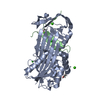
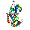
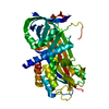
| Structure viewer | Molecule: MolmilJmol/JSmol |
|---|
- Downloads & links
-Download
| PDBx/mmCIF format | 1by7.cif.gz | 88.6 KB | Display | PDBx/mmCIF format |
|---|---|---|---|---|
| PDB format | pdb1by7.ent.gz | 65.3 KB | Display | PDB format |
| PDBx/mmJSON format | 1by7.json.gz | Tree view | PDBx/mmJSON format | |
| Others |  Other downloads Other downloads |
-Validation report
| Arichive directory | https://data.pdbj.org/pub/pdb/validation_reports/by/1by7ftp://data.pdbj.org/pub/pdb/validation_reports/by/1by7 | HTTPS FTP |
|---|
-Related structure data
| Similar structure data |
|---|
-Links
 PDBj
PDBj


- Assembly
| Deposited unit | 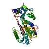
| ||||||||
|---|---|---|---|---|---|---|---|---|---|
| 1 |
| ||||||||
| Unit cell |
|
-Components
| #1: Protein | Mass: 43045.020 Da / Num. of mol.: 1 / Mutation: RESIDUES 66 - 98 EXCISED Source method: isolated from a genetically manipulated source Source: (gene. exp.) Homo sapiens (human) / Production host:  |
|---|---|
| #2: Water | ChemComp-HOH /  Mass: 18.015 Da / Num. of mol.: 209 / Source method: isolated from a natural source / Formula: H2O Mass: 18.015 Da / Num. of mol.: 209 / Source method: isolated from a natural source / Formula: H2O |
| Has protein modification | Y |
-Experimental details
-Experiment
| Experiment | Method: X-RAY DIFFRACTION / Number of used crystals: 1 |
|---|
- Sample preparation
| Crystal | Density Matthews: 2.36 Å3/Da / Density % sol: 47.92 % | ||||||||||||||||||||||||||||||
|---|---|---|---|---|---|---|---|---|---|---|---|---|---|---|---|---|---|---|---|---|---|---|---|---|---|---|---|---|---|---|---|
| Crystal grow | pH: 7.5 / Details: pH 7.5 | ||||||||||||||||||||||||||||||
| Crystal grow | *PLUS Method: vapor diffusion | ||||||||||||||||||||||||||||||
| Components of the solutions | *PLUS
|
-Data collection
| Diffraction | Mean temperature: 100 K |
|---|---|
| Diffraction source | Source: SYNCHROTRON / Site: SSRL  / Beamline: BL7-1 / Wavelength: 1.08 / Beamline: BL7-1 / Wavelength: 1.08 |
| Detector | Type: MARRESEARCH / Detector: IMAGE PLATE / Date: Jun 15, 1996 |
| Radiation | Protocol: SINGLE WAVELENGTH / Monochromatic (M) / Laue (L): M / Scattering type: x-ray |
| Radiation wavelength | Wavelength: 1.08 Å / Relative weight: 1 |
| Reflection | Resolution: 2→30 Å / Num. obs: 27274 / % possible obs: 99 % / Observed criterion σ(I): 0 / Redundancy: 6.4 % / Rmerge(I) obs: 0.055 / Rsym value: 0.055 |
| Reflection shell | Resolution: 2→2.1 Å / Redundancy: 5.8 % / Rmerge(I) obs: 0.35 / Rsym value: 0.35 / % possible all: 98 |
| Reflection | *PLUS Num. measured all: 176011 |
| Reflection shell | *PLUS % possible obs: 90.5 % |
- Processing
| Software |
| |||||||||||||||||||||||||||||||||||||||||||||||||||||||||||||||
|---|---|---|---|---|---|---|---|---|---|---|---|---|---|---|---|---|---|---|---|---|---|---|---|---|---|---|---|---|---|---|---|---|---|---|---|---|---|---|---|---|---|---|---|---|---|---|---|---|---|---|---|---|---|---|---|---|---|---|---|---|---|---|---|---|
| Refinement | Method to determine structure: MIR / Resolution: 2→30 Å / Cross valid method: THROUGHOUT / σ(F): 0
| |||||||||||||||||||||||||||||||||||||||||||||||||||||||||||||||
| Refinement step | Cycle: LAST / Resolution: 2→30 Å
| |||||||||||||||||||||||||||||||||||||||||||||||||||||||||||||||
| Refine LS restraints |
| |||||||||||||||||||||||||||||||||||||||||||||||||||||||||||||||
| Software | *PLUS Name: REFMAC / Classification: refinement | |||||||||||||||||||||||||||||||||||||||||||||||||||||||||||||||
| Refinement | *PLUS Highest resolution: 2 Å / σ(F): 0 / % reflection Rfree: 5 % / Rfactor obs: 0.202 | |||||||||||||||||||||||||||||||||||||||||||||||||||||||||||||||
| Solvent computation | *PLUS | |||||||||||||||||||||||||||||||||||||||||||||||||||||||||||||||
| Displacement parameters | *PLUS | |||||||||||||||||||||||||||||||||||||||||||||||||||||||||||||||
| Refine LS restraints | *PLUS Type: p_angle_d / Dev ideal: 0.04 |
