 Movie
Movie Controller


+ Open data

- Basic information
| Entry | Database: PDB / ID: 1i8d | ||||||
|---|---|---|---|---|---|---|---|
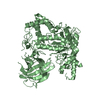
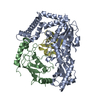
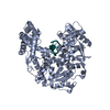
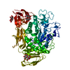
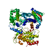
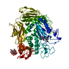
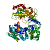
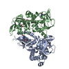
| Title | CRYSTAL STRUCTURE OF RIBOFLAVIN SYNTHASE | ||||||
 Components Components | RIBOFLAVIN SYNTHASE | ||||||
 Keywords Keywords | TRANSFERASE / riboflavin synthase / riboflavin biosynthesis / antimicrobial target / structure-based design / Escherichia coli | ||||||
| Function / homology |  Function and homology information Function and homology informationriboflavin synthase / riboflavin synthase activity / riboflavin biosynthetic process / cytosol Similarity search - Function | ||||||
| Biological species |  | ||||||
| Method |  X-RAY DIFFRACTION / SYNCHROTRON / MAD / Resolution: 2 Å X-RAY DIFFRACTION / SYNCHROTRON / MAD / Resolution: 2 Å | ||||||
 Authors Authors | Liao, D.-I. / Wawrzak, Z. / Calabrese, J.C. / Viitanen, P.V. / Jordan, D.B. | ||||||
 Citation Citation | Journal: Structure / Year: 2001 Title: Crystal structure of riboflavin synthase. Authors: Liao, D.I. / Wawrzak, Z. / Calabrese, J.C. / Viitanen, P.V. / Jordan, D.B. | ||||||
| History |
|
- Structure visualization
| Structure viewer | Molecule: MolmilJmol/JSmol |
|---|
- Downloads & links
-Download
| PDBx/mmCIF format | 1i8d.cif.gz | 127.2 KB | Display | PDBx/mmCIF format |
|---|---|---|---|---|
| PDB format | pdb1i8d.ent.gz | 100.4 KB | Display | PDB format |
| PDBx/mmJSON format | 1i8d.json.gz | Tree view | PDBx/mmJSON format | |
| Others |  Other downloads Other downloads |
-Validation report
| Summary document | 1i8d_validation.pdf.gz | 388.3 KB | Display | wwPDB validaton report |
|---|---|---|---|---|
| Full document | 1i8d_full_validation.pdf.gz | 417.9 KB | Display | |
| Data in XML | 1i8d_validation.xml.gz | 15.7 KB | Display | |
| Data in CIF | 1i8d_validation.cif.gz | 24.7 KB | Display | |
| Arichive directory | https://data.pdbj.org/pub/pdb/validation_reports/i8/1i8dftp://data.pdbj.org/pub/pdb/validation_reports/i8/1i8d | HTTPS FTP |
-Related structure data
| Similar structure data |
|---|


-Links
 PDBj
PDBj


- Assembly
| Deposited unit | 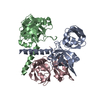
| ||||||||
|---|---|---|---|---|---|---|---|---|---|
| 1 |
| ||||||||
| Unit cell |
| ||||||||
| Details | The biological assembly is a homo trimer. The three molecules in the asymmetric unit represent the biological assembly. The trimer is asymmetrical. |
-Components
| #1: Protein | Mass: 23472.885 Da / Num. of mol.: 3 Source method: isolated from a genetically manipulated source Source: (gene. exp.) References: UniProt: P29015, UniProt: P0AFU8*PLUS, riboflavin synthase #2: Water | ChemComp-HOH / |  Mass: 18.015 Da / Num. of mol.: 229 / Source method: isolated from a natural source / Formula: H2O Mass: 18.015 Da / Num. of mol.: 229 / Source method: isolated from a natural source / Formula: H2O |
|---|
-Experimental details
-Experiment
| Experiment | Method: X-RAY DIFFRACTION / Number of used crystals: 1 |
|---|
- Sample preparation
| Crystal | Density Matthews: 2.51 Å3/Da / Density % sol: 50.94 % | ||||||||||||||||||||||||||||||||||||||||||
|---|---|---|---|---|---|---|---|---|---|---|---|---|---|---|---|---|---|---|---|---|---|---|---|---|---|---|---|---|---|---|---|---|---|---|---|---|---|---|---|---|---|---|---|
| Crystal grow | Temperature: 295 K / Method: vapor diffusion, hanging drop / pH: 7.5 Details: bis-tris propane, MgCl2, diglyme, pH 7.5, VAPOR DIFFUSION, HANGING DROP, temperature 295K | ||||||||||||||||||||||||||||||||||||||||||
| Crystal grow | *PLUS Temperature: 22 ℃ / Method: vapor diffusion, sitting drop | ||||||||||||||||||||||||||||||||||||||||||
| Components of the solutions | *PLUS
|
-Data collection
| Diffraction | Mean temperature: 93 K |
|---|---|
| Diffraction source | Source: SYNCHROTRON / Site: APS  / Beamline: 5ID-B / Wavelength: 1.1271 Å / Beamline: 5ID-B / Wavelength: 1.1271 Å |
| Detector | Type: MARRESEARCH / Detector: CCD / Date: Jan 1, 1999 |
| Radiation | Protocol: SINGLE WAVELENGTH / Monochromatic (M) / Laue (L): M / Scattering type: x-ray |
| Radiation wavelength | Wavelength: 1.1271 Å / Relative weight: 1 |
| Reflection | Resolution: 2→25 Å / Num. all: 48686 / Num. obs: 437196 / % possible obs: 99.2 % / Observed criterion σ(I): -3 / Redundancy: 9 % / Rmerge(I) obs: 0.064 / Net I/σ(I): 33.7 |
| Reflection shell | Resolution: 2→2.04 Å / Redundancy: 5 % / Rmerge(I) obs: 0.215 / % possible all: 96.7 |
| Reflection | *PLUS Num. obs: 48686 / Num. measured all: 437196 |
| Reflection shell | *PLUS % possible obs: 96.7 % / Mean I/σ(I) obs: 5.6 |
- Processing
| Software |
| |||||||||||||||
|---|---|---|---|---|---|---|---|---|---|---|---|---|---|---|---|---|
| Refinement | Method to determine structure: MAD / Resolution: 2→25 Å / σ(F): 2 / Stereochemistry target values: TNT library
| |||||||||||||||
| Refinement step | Cycle: LAST / Resolution: 2→25 Å
| |||||||||||||||
| Refine LS restraints |
| |||||||||||||||
| Software | *PLUS Name: TNT / Version: 5E / Classification: refinement | |||||||||||||||
| Refinement | *PLUS Highest resolution: 2 Å / Lowest resolution: 25 Å / σ(F): 2 / % reflection Rfree: 5 % / Rfactor obs: 0.239 | |||||||||||||||
| Solvent computation | *PLUS | |||||||||||||||
| Displacement parameters | *PLUS | |||||||||||||||
| Refine LS restraints | *PLUS Type: t_angle_deg / Dev ideal: 1.99 |
