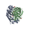
 Movie
Movie Controller

[English] 日本語
 Yorodumi
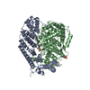
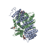
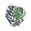
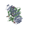
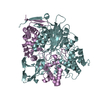
- PDB-1l1l: CRYSTAL STRUCTURE OF B-12 DEPENDENT (CLASS II) RIBONUCLEOTIDE RED... -
+ Open data

- Basic information
| Entry | Database: PDB / ID: 1l1l | ||||||
|---|---|---|---|---|---|---|---|
| Title | CRYSTAL STRUCTURE OF B-12 DEPENDENT (CLASS II) RIBONUCLEOTIDE REDUCTASE | ||||||
 Components Components | RIBONUCLEOSIDE TRIPHOSPHATE REDUCTASE | ||||||
 Keywords Keywords | OXIDOREDUCTASE / 10-stranded alpha-beta barrel / central finger loop | ||||||
| Function / homology |  Function and homology information Function and homology informationribonucleoside-triphosphate reductase (thioredoxin) / ribonucleoside-triphosphate reductase (thioredoxin) activity / cobalamin binding / ribonucleoside-diphosphate reductase activity, thioredoxin disulfide as acceptor / DNA replication / nucleotide binding Similarity search - Function | ||||||
| Biological species |  Lactobacillus leichmannii (bacteria) Lactobacillus leichmannii (bacteria) | ||||||
| Method |  X-RAY DIFFRACTION / SYNCHROTRON / MAD / Resolution: 1.75 Å X-RAY DIFFRACTION / SYNCHROTRON / MAD / Resolution: 1.75 Å | ||||||
 Authors Authors | Sintchak, M.D. / Arjara, G. / Kellogg, B.A. / Stubbe, J. / Drennan, C.L. | ||||||
 Citation Citation | Journal: Nat.Struct.Biol. / Year: 2002 Title: The crystal structure of class II ribonucleotide reductase reveals how an allosterically regulated monomer mimics a dimer. Authors: Sintchak, M.D. / Arjara, G. / Kellogg, B.A. / Stubbe, J. / Drennan, C.L. | ||||||
| History |
| ||||||
| Remark 999 | SEQUENCE Author states the sequence is correct and residue number 152 is PHE, there is an error in ...SEQUENCE Author states the sequence is correct and residue number 152 is PHE, there is an error in the reference database. |
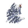
- Structure visualization
| Structure viewer | Molecule: MolmilJmol/JSmol |
|---|
- Downloads & links
-Download
| PDBx/mmCIF format | 1l1l.cif.gz | 612.5 KB | Display | PDBx/mmCIF format |
|---|---|---|---|---|
| PDB format | pdb1l1l.ent.gz | 499.8 KB | Display | PDB format |
| PDBx/mmJSON format | 1l1l.json.gz | Tree view | PDBx/mmJSON format | |
| Others |  Other downloads Other downloads |
-Validation report
| Arichive directory | https://data.pdbj.org/pub/pdb/validation_reports/l1/1l1lftp://data.pdbj.org/pub/pdb/validation_reports/l1/1l1l | HTTPS FTP |
|---|
-Related structure data
| Similar structure data |
|---|

-Links
 PDBj
PDBj
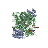
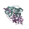
- Assembly
| Deposited unit | 
| ||||||||
|---|---|---|---|---|---|---|---|---|---|
| 1 | 
| ||||||||
| 2 | 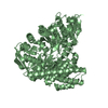
| ||||||||
| 3 | 
| ||||||||
| 4 | 
| ||||||||
| Unit cell |
| ||||||||
| Details | the biological assembly is a monomer |
-Components
| #1: Protein | Mass: 82070.969 Da / Num. of mol.: 4 / Mutation: C731/736S Source method: isolated from a genetically manipulated source Source: (gene. exp.) Lactobacillus leichmannii (bacteria) / Production host: References: UniProt: Q59490, ribonucleoside-triphosphate reductase (thioredoxin) #2: Water | ChemComp-HOH / |  Mass: 18.015 Da / Num. of mol.: 2875 / Source method: isolated from a natural source / Formula: H2O Mass: 18.015 Da / Num. of mol.: 2875 / Source method: isolated from a natural source / Formula: H2OHas protein modification | Y | |
|---|
-Experimental details
-Experiment
| Experiment | Method: X-RAY DIFFRACTION / Number of used crystals: 1 |
|---|
- Sample preparation
| Crystal | Density Matthews: 2.4 Å3/Da / Density % sol: 48.65 % | ||||||||||||||||||||||||||||||||||||||||||||||||||||||||||||
|---|---|---|---|---|---|---|---|---|---|---|---|---|---|---|---|---|---|---|---|---|---|---|---|---|---|---|---|---|---|---|---|---|---|---|---|---|---|---|---|---|---|---|---|---|---|---|---|---|---|---|---|---|---|---|---|---|---|---|---|---|---|
| Crystal grow | Temperature: 298 K / Method: vapor diffusion, hanging drop / pH: 5.6 Details: ammonium sulfate, PEG 8000, glycerol, sodium citrate, pH 5.6, VAPOR DIFFUSION, HANGING DROP, temperature 298K | ||||||||||||||||||||||||||||||||||||||||||||||||||||||||||||
| Crystal grow | *PLUS | ||||||||||||||||||||||||||||||||||||||||||||||||||||||||||||
| Components of the solutions | *PLUS
|
-Data collection
| Diffraction | Mean temperature: 100 K |
|---|---|
| Diffraction source | Source: SYNCHROTRON / Site: NSLS  / Beamline: X25 / Wavelength: 1.1022 Å / Beamline: X25 / Wavelength: 1.1022 Å |
| Detector | Type: BRANDEIS - B4 / Detector: CCD / Date: Jul 28, 2000 |
| Radiation | Monochromator: graphite / Protocol: SINGLE WAVELENGTH / Monochromatic (M) / Laue (L): M / Scattering type: x-ray |
| Radiation wavelength | Wavelength: 1.1022 Å / Relative weight: 1 |
| Reflection | Resolution: 1.75→30 Å / Num. all: 285688 / Num. obs: 285688 / % possible obs: 91.6 % / Observed criterion σ(F): 0 / Observed criterion σ(I): 0 / Redundancy: 2.2 % / Rsym value: 0.058 / Net I/σ(I): 13.5 |
| Reflection shell | Resolution: 1.75→1.81 Å / Mean I/σ(I) obs: 2.1 / Rsym value: 0.267 / % possible all: 60.2 |
| Reflection | *PLUS Lowest resolution: 30 Å / Num. measured all: 632497 / Rmerge(I) obs: 0.058 |
| Reflection shell | *PLUS % possible obs: 60.2 % / Rmerge(I) obs: 0.267 |
- Processing
| Software |
| ||||||||||||||||||||
|---|---|---|---|---|---|---|---|---|---|---|---|---|---|---|---|---|---|---|---|---|---|
| Refinement | Method to determine structure: MAD / Resolution: 1.75→30 Å / Cross valid method: THROUGHOUT / σ(F): 1 / Stereochemistry target values: Engh & Huber
| ||||||||||||||||||||
| Refinement step | Cycle: LAST / Resolution: 1.75→30 Å
| ||||||||||||||||||||
| Refine LS restraints |
| ||||||||||||||||||||
| LS refinement shell | Resolution: 1.75→1.76 Å
| ||||||||||||||||||||
| Refinement | *PLUS Lowest resolution: 500 Å / Num. reflection obs: 272996 / Rfactor obs: 0.223 | ||||||||||||||||||||
| Solvent computation | *PLUS | ||||||||||||||||||||
| Displacement parameters | *PLUS | ||||||||||||||||||||
| Refine LS restraints | *PLUS
|
