 Movie
Movie Controller


+ Open data

- Basic information
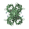
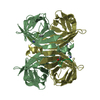
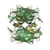
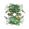
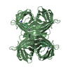
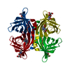
| Entry | Database: PDB / ID: 1hxz | ||||||
|---|---|---|---|---|---|---|---|
| Title | MINIPROTEIN MP-2 COMPLEX WITH STREPTAVIDIN | ||||||
 Components Components |
| ||||||
 Keywords Keywords | UNKNOWN FUNCTION / CONFORMATIONAL ENSEMBLE / MINI-PROTEINS / DISULPHIDE CONSTRAINED LOOPS / ENTROPICALLY RESTRAINED PROTEINS / PEPTIDES | ||||||
| Function / homology |  Function and homology information Function and homology information | ||||||
| Biological species |  Streptomyces avidinii (bacteria) Streptomyces avidinii (bacteria) | ||||||
| Method |  X-RAY DIFFRACTION / MOLECULAR REPLACEMENT / Resolution: 1.8 Å X-RAY DIFFRACTION / MOLECULAR REPLACEMENT / Resolution: 1.8 Å | ||||||
 Authors Authors | Yang, H.W. / Liu, D.Q. / Fan, X. / White, M.A. / Fox, R.O. | ||||||
 Citation Citation | Journal: To be Published Title: Conformational Ensemble Analysis of Ligand Binding in Streptavidin Mini-Protein Complexes Authors: Yang, H.W. / Liu, D.Q. / Fan, X. / White, M.A. / Fox, R.O. | ||||||
| History |
|
- Structure visualization
| Structure viewer | Molecule: MolmilJmol/JSmol |
|---|
- Downloads & links
-Download
| PDBx/mmCIF format | 1hxz.cif.gz | 64.4 KB | Display | PDBx/mmCIF format |
|---|---|---|---|---|
| PDB format | pdb1hxz.ent.gz | 47.6 KB | Display | PDB format |
| PDBx/mmJSON format | 1hxz.json.gz | Tree view | PDBx/mmJSON format | |
| Others |  Other downloads Other downloads |
-Validation report
| Summary document | 1hxz_validation.pdf.gz | 454.4 KB | Display | wwPDB validaton report |
|---|---|---|---|---|
| Full document | 1hxz_full_validation.pdf.gz | 458.5 KB | Display | |
| Data in XML | 1hxz_validation.xml.gz | 13.7 KB | Display | |
| Data in CIF | 1hxz_validation.cif.gz | 18.8 KB | Display | |
| Arichive directory | https://data.pdbj.org/pub/pdb/validation_reports/hx/1hxzftp://data.pdbj.org/pub/pdb/validation_reports/hx/1hxz | HTTPS FTP |
-Related structure data
| Related structure data |  1hxlC  1hy2C  1sldS C: citing same article ( S: Starting model for refinement |
|---|---|
| Similar structure data |


-Links
 PDBj
PDBj- Assembly
| Deposited unit | 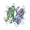
| ||||||||
|---|---|---|---|---|---|---|---|---|---|
| 1 | 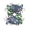
| ||||||||
| Unit cell |
| ||||||||
| Details | The second part of the biological assembly is generated from the dimer by the two fold axis: -x,y,1/2-z |
-Components
| #1: Protein | Mass: 13409.466 Da / Num. of mol.: 2 Source method: isolated from a genetically manipulated source Source: (gene. exp.) Streptomyces avidinii (bacteria) / Production host: #2: Protein/peptide | Mass: 1653.970 Da / Num. of mol.: 2 / Source method: obtained synthetically / Details: THE PEPTIDE WAS CHEMICALLY SYNTHESIZED. #3: Water | ChemComp-HOH / |  Mass: 18.015 Da / Num. of mol.: 162 / Source method: isolated from a natural source / Formula: H2O Mass: 18.015 Da / Num. of mol.: 162 / Source method: isolated from a natural source / Formula: H2OHas protein modification | Y | |
|---|
-Experimental details
-Experiment
| Experiment | Method: X-RAY DIFFRACTION / Number of used crystals: 1 |
|---|
- Sample preparation
| Crystal | Density Matthews: 2.51 Å3/Da / Density % sol: 50.95 % |
|---|---|
| Crystal grow | Temperature: 298 K / Method: vapor diffusion, hanging drop / pH: 4 Details: 100mM potassium acetate, 32% AMMONIUM SULFATE , pH 4.0, VAPOR DIFFUSION, HANGING DROP, temperature 298K |
-Data collection
| Diffraction | Mean temperature: 277 K |
|---|---|
| Diffraction source | Source: ROTATING ANODE / Type: MACSCIENCE / Wavelength: 1.5418 |
| Detector | Type: MACSCIENCE / Detector: IMAGE PLATE / Date: Jan 14, 1999 / Details: MULTILAYER |
| Radiation | Monochromator: MULTILAYER OPTICS / Protocol: SINGLE WAVELENGTH / Monochromatic (M) / Laue (L): M / Scattering type: x-ray |
| Radiation wavelength | Wavelength: 1.5418 Å / Relative weight: 1 |
| Reflection | Resolution: 1.8→29.1 Å / Num. obs: 28243 / % possible obs: 99.2 % / Observed criterion σ(I): 0 / Redundancy: 4.87 % / Biso Wilson estimate: 8.4 Å2 / Rmerge(I) obs: 0.061 / Rsym value: 0.061 / Net I/σ(I): 12.9 |
| Reflection shell | Resolution: 1.78→1.81 Å / Redundancy: 4.5 % / Rmerge(I) obs: 0.134 / Rsym value: 0.134 / % possible all: 98.7 |
- Processing
| Software |
| ||||||||||||||||||||||||||||||||||||||||||||||||||||||||||||||||||||||||||||||||
|---|---|---|---|---|---|---|---|---|---|---|---|---|---|---|---|---|---|---|---|---|---|---|---|---|---|---|---|---|---|---|---|---|---|---|---|---|---|---|---|---|---|---|---|---|---|---|---|---|---|---|---|---|---|---|---|---|---|---|---|---|---|---|---|---|---|---|---|---|---|---|---|---|---|---|---|---|---|---|---|---|---|
| Refinement | Method to determine structure: MOLECULAR REPLACEMENT Starting model: 1SLD STREPTAVIDIN TETRAMER Resolution: 1.8→29.1 Å / Rfactor Rfree error: 0.004 / Data cutoff high absF: 747543.62 / Data cutoff low absF: 0 / Isotropic thermal model: RESTRAINED / Cross valid method: THROUGHOUT / σ(F): 0 / Stereochemistry target values: Engh & Huber
| ||||||||||||||||||||||||||||||||||||||||||||||||||||||||||||||||||||||||||||||||
| Solvent computation | Solvent model: FLAT MODEL / Bsol: 39.54 Å2 / ksol: 0.331 e/Å3 | ||||||||||||||||||||||||||||||||||||||||||||||||||||||||||||||||||||||||||||||||
| Displacement parameters | Biso mean: 18.4 Å2
| ||||||||||||||||||||||||||||||||||||||||||||||||||||||||||||||||||||||||||||||||
| Refine analyze |
| ||||||||||||||||||||||||||||||||||||||||||||||||||||||||||||||||||||||||||||||||
| Refinement step | Cycle: LAST / Resolution: 1.8→29.1 Å
| ||||||||||||||||||||||||||||||||||||||||||||||||||||||||||||||||||||||||||||||||
| Refine LS restraints |
| ||||||||||||||||||||||||||||||||||||||||||||||||||||||||||||||||||||||||||||||||
| LS refinement shell | Resolution: 1.8→1.91 Å / Rfactor Rfree error: 0.012 / Total num. of bins used: 6
| ||||||||||||||||||||||||||||||||||||||||||||||||||||||||||||||||||||||||||||||||
| Xplor file |
|
