 Movie
Movie Controller


+ Open data

- Basic information
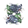
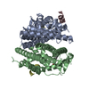
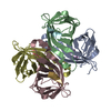
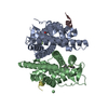
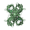
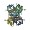
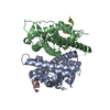
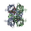
| Entry | Database: PDB / ID: 1hqq | ||||||
|---|---|---|---|---|---|---|---|
| Title | MINIPROTEIN MP-2 (M9A) COMPLEX WITH STREPTAVIDIN | ||||||
 Components Components |
| ||||||
 Keywords Keywords | UNKNOWN FUNCTION / Conformational Ensemble / mini-proteins / Disulphide constrained loops / Entropically restrained proteins / peptides | ||||||
| Function / homology |  Function and homology information Function and homology information | ||||||
| Biological species |  Streptomyces avidinii (bacteria) Streptomyces avidinii (bacteria) | ||||||
| Method |  X-RAY DIFFRACTION / MOLECULAR REPLACEMENT / Resolution: 1.7 Å X-RAY DIFFRACTION / MOLECULAR REPLACEMENT / Resolution: 1.7 Å | ||||||
 Authors Authors | Yang, H.W. / Liu, D.Q. / Fan, X. / White, M.A. / Fox, R.O. | ||||||
 Citation Citation | Journal: To be Published Title: Conformational Ensemble Analysis of Ligand Binding in Streptavidin Mini-protein Complexes Authors: Yang, H.W. / Liu, D.Q. / Fan, X. / White, M.A. / Fox, R.O. | ||||||
| History |
|
- Structure visualization
| Structure viewer | Molecule: MolmilJmol/JSmol |
|---|
- Downloads & links
-Download
| PDBx/mmCIF format | 1hqq.cif.gz | 110.3 KB | Display | PDBx/mmCIF format |
|---|---|---|---|---|
| PDB format | pdb1hqq.ent.gz | 85.1 KB | Display | PDB format |
| PDBx/mmJSON format | 1hqq.json.gz | Tree view | PDBx/mmJSON format | |
| Others |  Other downloads Other downloads |
-Validation report
| Arichive directory | https://data.pdbj.org/pub/pdb/validation_reports/hq/1hqqftp://data.pdbj.org/pub/pdb/validation_reports/hq/1hqq | HTTPS FTP |
|---|
-Related structure data
| Related structure data | |
|---|---|
| Similar structure data |


-Links
 PDBj
PDBj- Assembly
| Deposited unit | 
| ||||||||
|---|---|---|---|---|---|---|---|---|---|
| 1 |
| ||||||||
| Unit cell |
|
-Components
| #1: Protein | Mass: 13409.466 Da / Num. of mol.: 4 Source method: isolated from a genetically manipulated source Source: (gene. exp.) Streptomyces avidinii (bacteria) / Production host: #2: Protein/peptide | Mass: 1593.852 Da / Num. of mol.: 4 / Mutation: M9A / Source method: obtained synthetically / Details: The peptide was chemically synthesized. #3: Water | ChemComp-HOH / |  Mass: 18.015 Da / Num. of mol.: 172 / Source method: isolated from a natural source / Formula: H2O Mass: 18.015 Da / Num. of mol.: 172 / Source method: isolated from a natural source / Formula: H2OHas protein modification | Y | |
|---|
-Experimental details
-Experiment
| Experiment | Method: X-RAY DIFFRACTION / Number of used crystals: 1 |
|---|
- Sample preparation
| Crystal | Density Matthews: 2.51 Å3/Da / Density % sol: 51.07 % |
|---|---|
| Crystal grow | Temperature: 298 K / Method: vapor diffusion, hanging drop / pH: 4 Details: 100 mM potassium acetate, ammonium sulfate, pH 4.0, VAPOR DIFFUSION, HANGING DROP, temperature 298K |
-Data collection
| Diffraction | Mean temperature: 108 K |
|---|---|
| Diffraction source | Source: ROTATING ANODE / Type: MACSCIENCE / Wavelength: 1.5418 Å |
| Detector | Type: MACSCIENCE / Detector: IMAGE PLATE / Date: Oct 20, 2000 / Details: Multilayer |
| Radiation | Monochromator: Multilayer optics / Protocol: SINGLE WAVELENGTH / Monochromatic (M) / Laue (L): M / Scattering type: x-ray |
| Radiation wavelength | Wavelength: 1.5418 Å / Relative weight: 1 |
| Reflection | Resolution: 1.7→20.84 Å / Num. all: 63318 / Num. obs: 163318 / % possible obs: 96.6 % / Observed criterion σ(F): 0 / Observed criterion σ(I): 0 / Redundancy: 4.46 % / Biso Wilson estimate: 12.5 Å2 / Limit h max: 34 / Limit h min: -34 / Limit k max: 52 / Limit k min: -34 / Limit l max: 33 / Limit l min: 0 / Observed criterion F max: 569614.15 / Observed criterion F min: 0.316 / Rmerge(I) obs: 0.086 / Rsym value: 0.086 / Net I/σ(I): 8.1 |
| Reflection shell | Resolution: 1.7→1.73 Å / Rmerge(I) obs: 0.302 / Mean I/σ(I) obs: 4.2 / Num. unique all: 2754 / Rsym value: 0.302 / % possible all: 84.8 |
- Processing
| Software |
| ||||||||||||||||||||||||||||||||||||||||||||||||||||||||||||||||||||||||||||||||||||||||||
|---|---|---|---|---|---|---|---|---|---|---|---|---|---|---|---|---|---|---|---|---|---|---|---|---|---|---|---|---|---|---|---|---|---|---|---|---|---|---|---|---|---|---|---|---|---|---|---|---|---|---|---|---|---|---|---|---|---|---|---|---|---|---|---|---|---|---|---|---|---|---|---|---|---|---|---|---|---|---|---|---|---|---|---|---|---|---|---|---|---|---|---|
| Refinement | Method to determine structure: MOLECULAR REPLACEMENT / Resolution: 1.7→20.84 Å / Rfactor Rfree error: 0.003 / Occupancy max: 1 / Occupancy min: 1 / Data cutoff high rms absF: 569571.22 / Isotropic thermal model: Isotropic / Cross valid method: THROUGHOUT / σ(F): 0 / σ(I): 0 / Stereochemistry target values: Engh & Huber
| ||||||||||||||||||||||||||||||||||||||||||||||||||||||||||||||||||||||||||||||||||||||||||
| Solvent computation | Solvent model: CNS bulk solvent model used / Bsol: 38.6719 Å2 / ksol: 0.351859 e/Å3 | ||||||||||||||||||||||||||||||||||||||||||||||||||||||||||||||||||||||||||||||||||||||||||
| Displacement parameters | Biso max: 48.9 Å2 / Biso mean: 17.08 Å2 / Biso min: 3.77 Å2
| ||||||||||||||||||||||||||||||||||||||||||||||||||||||||||||||||||||||||||||||||||||||||||
| Refine analyze |
| ||||||||||||||||||||||||||||||||||||||||||||||||||||||||||||||||||||||||||||||||||||||||||
| Refinement step | Cycle: LAST / Resolution: 1.7→20.84 Å
| ||||||||||||||||||||||||||||||||||||||||||||||||||||||||||||||||||||||||||||||||||||||||||
| Refine LS restraints |
| ||||||||||||||||||||||||||||||||||||||||||||||||||||||||||||||||||||||||||||||||||||||||||
| LS refinement shell | Refine-ID: X-RAY DIFFRACTION / Total num. of bins used: 8
| ||||||||||||||||||||||||||||||||||||||||||||||||||||||||||||||||||||||||||||||||||||||||||
| Xplor file |
|
