 Movie
Movie Controller

[English] 日本語
 Yorodumi
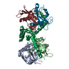
- PDB-1hoz: CRYSTAL STRUCTURE OF AN INOSINE-ADENOSINE-GUANOSINE-PREFERRING NU... -
+ Open data

- Basic information
| Entry | Database: PDB / ID: 1hoz | ||||||
|---|---|---|---|---|---|---|---|
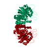
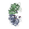
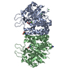
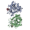
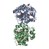
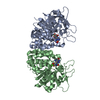
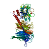
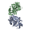
| Title | CRYSTAL STRUCTURE OF AN INOSINE-ADENOSINE-GUANOSINE-PREFERRING NUCLEOSIDE HYDROLASE FROM TRYPANOSOMA VIVAX | ||||||
 Components Components | INOSINE-ADENOSINE-GUANOSINE-PREFERRING NUCLEOSIDE HYDROLASE | ||||||
 Keywords Keywords | HYDROLASE / Rossmann-fold-like motif | ||||||
| Function / homology |  Function and homology information Function and homology informationpurine nucleosidase activity / purine nucleoside catabolic process / metal ion binding / cytosol Similarity search - Function | ||||||
| Biological species |  | ||||||
| Method |  X-RAY DIFFRACTION / SYNCHROTRON / MAD / Resolution: 1.6 Å X-RAY DIFFRACTION / SYNCHROTRON / MAD / Resolution: 1.6 Å | ||||||
 Authors Authors | Versees, W. / Decanniere, K. / Pelle, R. / Depoorter, J. / Parkin, D.W. / Steyaert, J. | ||||||
 Citation Citation | Journal: J.Mol.Biol. / Year: 2001 Title: Structure and function of a novel purine specific nucleoside hydrolase from Trypanosoma vivax. Authors: Versees, W. / Decanniere, K. / Pelle, R. / Depoorter, J. / Brosens, E. / Parkin, D.W. / Steyaert, J. | ||||||
| History |
|
- Structure visualization
| Structure viewer | Molecule: MolmilJmol/JSmol |
|---|
- Downloads & links
-Download
| PDBx/mmCIF format | 1hoz.cif.gz | 146.6 KB | Display | PDBx/mmCIF format |
|---|---|---|---|---|
| PDB format | pdb1hoz.ent.gz | 114.6 KB | Display | PDB format |
| PDBx/mmJSON format | 1hoz.json.gz | Tree view | PDBx/mmJSON format | |
| Others |  Other downloads Other downloads |
-Validation report
| Arichive directory | https://data.pdbj.org/pub/pdb/validation_reports/ho/1hozftp://data.pdbj.org/pub/pdb/validation_reports/ho/1hoz | HTTPS FTP |
|---|
-Related structure data

-Links
 PDBj
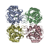
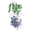
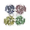
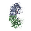
PDBj- Assembly
| Deposited unit | 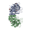
| ||||||||
|---|---|---|---|---|---|---|---|---|---|
| 1 |
| ||||||||
| Unit cell |
|
-Components
| #1: Protein | Mass: 37718.984 Da / Num. of mol.: 2 / Fragment: IAG-NUCLEOSIDE HYDROLASE, IAG-NH Source method: isolated from a genetically manipulated source Source: (gene. exp.)  #2: Chemical |   Mass: 40.078 Da / Num. of mol.: 2 / Source method: obtained synthetically / Formula: Ca Mass: 40.078 Da / Num. of mol.: 2 / Source method: obtained synthetically / Formula: Ca#3: Chemical | ChemComp-GOL /   Mass: 92.094 Da / Num. of mol.: 8 / Source method: obtained synthetically / Formula: C3H8O3 Mass: 92.094 Da / Num. of mol.: 8 / Source method: obtained synthetically / Formula: C3H8O3#4: Water | ChemComp-HOH / |  Mass: 18.015 Da / Num. of mol.: 424 / Source method: isolated from a natural source / Formula: H2O Mass: 18.015 Da / Num. of mol.: 424 / Source method: isolated from a natural source / Formula: H2O |
|---|
-Experimental details
-Experiment
| Experiment | Method: X-RAY DIFFRACTION / Number of used crystals: 1 |
|---|
- Sample preparation
| Crystal | Density Matthews: 2.03 Å3/Da / Density % sol: 39.31 % | ||||||||||||||||||||||||
|---|---|---|---|---|---|---|---|---|---|---|---|---|---|---|---|---|---|---|---|---|---|---|---|---|---|
| Crystal grow | Temperature: 293 K / Method: vapor diffusion, hanging drop / pH: 8.5 Details: 100 mM tris, 1.6 M ammonium sulfate, pH 8.5, VAPOR DIFFUSION, HANGING DROP, temperature 293K | ||||||||||||||||||||||||
| Crystal grow | *PLUS | ||||||||||||||||||||||||
| Components of the solutions | *PLUS
|
-Data collection
| Diffraction | Mean temperature: 100 K |
|---|---|
| Diffraction source | Source: SYNCHROTRON / Site: EMBL/DESY, HAMBURG  / Beamline: BW7B / Wavelength: 0.8445 Å / Beamline: BW7B / Wavelength: 0.8445 Å |
| Detector | Type: MARRESEARCH / Detector: IMAGE PLATE / Date: Nov 10, 1999 |
| Radiation | Monochromator: YALE MIRRORS / Protocol: MAD / Monochromatic (M) / Laue (L): M / Scattering type: x-ray |
| Radiation wavelength | Wavelength: 0.8445 Å / Relative weight: 1 |
| Reflection | Resolution: 1.6→30 Å / Num. all: 631096 / Num. obs: 629834 / % possible obs: 99.8 % / Observed criterion σ(F): 0 / Observed criterion σ(I): 0 / Redundancy: 4.4 % / Biso Wilson estimate: 16.8 Å2 / Rmerge(I) obs: 0.055 / Net I/σ(I): 10.4 |
| Reflection shell | Resolution: 1.6→1.69 Å / Redundancy: 3.4 % / Rmerge(I) obs: 0.38 / Mean I/σ(I) obs: 2 / % possible all: 99.5 |
| Reflection | *PLUS Num. measured all: 629834 |
| Reflection shell | *PLUS % possible obs: 99.5 % / Rmerge(I) obs: 0.385 |
- Processing
| Software |
| ||||||||||||||||||||
|---|---|---|---|---|---|---|---|---|---|---|---|---|---|---|---|---|---|---|---|---|---|
| Refinement | Method to determine structure: MAD / Resolution: 1.6→30 Å / σ(F): 0 / σ(I): 0 / Stereochemistry target values: Engh & Huber
| ||||||||||||||||||||
| Refinement step | Cycle: LAST / Resolution: 1.6→30 Å
| ||||||||||||||||||||
| Refine LS restraints |
| ||||||||||||||||||||
| LS refinement shell |
| ||||||||||||||||||||
| Software | *PLUS Name: CNS / Classification: refinement | ||||||||||||||||||||
| Refinement | *PLUS Highest resolution: 1.6 Å / Lowest resolution: 30 Å / σ(F): 0 / Rfactor obs: 0.1861 / Rfactor Rfree: 0.2096 | ||||||||||||||||||||
| Solvent computation | *PLUS | ||||||||||||||||||||
| Displacement parameters | *PLUS | ||||||||||||||||||||
| LS refinement shell | *PLUS Rfactor Rfree: 0.184 / Rfactor Rwork: 0.169 |
