 Movie
Movie Controller

[English] 日本語
 Yorodumi
- PDB-1kic: Inosine-adenosine-guanosine preferring nucleoside hydrolase from ... -
+ Open data

- Basic information
| Entry | Database: PDB / ID: 1kic | ||||||
|---|---|---|---|---|---|---|---|
| Title | Inosine-adenosine-guanosine preferring nucleoside hydrolase from Trypanosoma vivax: Asp10Ala mutant in complex with inosine | ||||||
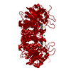
 Components Components | inosine-adenosine-guanosine preferring nucleoside hydrolase | ||||||
 Keywords Keywords | HYDROLASE / Rossmann-fold-like motif | ||||||
| Function / homology |  Function and homology information Function and homology informationpurine nucleosidase activity / purine nucleoside catabolic process / metal ion binding / cytosol Similarity search - Function | ||||||
| Biological species |  | ||||||
| Method |  X-RAY DIFFRACTION / SYNCHROTRON / MOLECULAR REPLACEMENT / Resolution: 1.6 Å X-RAY DIFFRACTION / SYNCHROTRON / MOLECULAR REPLACEMENT / Resolution: 1.6 Å | ||||||
 Authors Authors | Versees, W. / Decanniere, K. / Van Holsbeke, E. / Devroede, N. / Steyaert, J. | ||||||
 Citation Citation | Journal: J.Biol.Chem. / Year: 2002 Title: Enzyme-substrate interactions in the purine-specific nucleoside hydrolase from Trypanosoma vivax. Authors: Versees, W. / Decanniere, K. / Van Holsbeke, E. / Devroede, N. / Steyaert, J. | ||||||
| History |
|
- Structure visualization
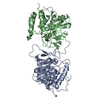
| Structure viewer | Molecule: MolmilJmol/JSmol |
|---|
- Downloads & links
-Download
| PDBx/mmCIF format | 1kic.cif.gz | 154.7 KB | Display | PDBx/mmCIF format |
|---|---|---|---|---|
| PDB format | pdb1kic.ent.gz | 118.2 KB | Display | PDB format |
| PDBx/mmJSON format | 1kic.json.gz | Tree view | PDBx/mmJSON format | |
| Others |  Other downloads Other downloads |
-Validation report
| Arichive directory | https://data.pdbj.org/pub/pdb/validation_reports/ki/1kicftp://data.pdbj.org/pub/pdb/validation_reports/ki/1kic | HTTPS FTP |
|---|
-Related structure data
| Related structure data |  1kieC  1hozS C: citing same article ( S: Starting model for refinement |
|---|---|
| Similar structure data |

-Links
 PDBj
PDBj- Assembly
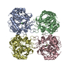
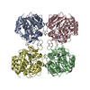
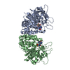
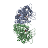
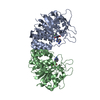
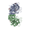
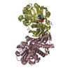
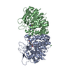
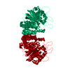
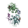
| Deposited unit | 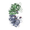
| ||||||||
|---|---|---|---|---|---|---|---|---|---|
| 1 |
| ||||||||
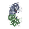
| Unit cell |
|
-Components
| #1: Protein | Mass: 37674.973 Da / Num. of mol.: 2 / Mutation: D10A Source method: isolated from a genetically manipulated source Source: (gene. exp.)  #2: Chemical |   Mass: 40.078 Da / Num. of mol.: 2 / Source method: obtained synthetically / Formula: Ca Mass: 40.078 Da / Num. of mol.: 2 / Source method: obtained synthetically / Formula: Ca#3: Chemical | ChemComp-NI / |   Mass: 58.693 Da / Num. of mol.: 1 / Source method: obtained synthetically / Formula: Ni Mass: 58.693 Da / Num. of mol.: 1 / Source method: obtained synthetically / Formula: Ni#4: Chemical | ChemComp-NOS / 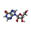  Mass: 268.226 Da / Num. of mol.: 4 / Source method: obtained synthetically / Formula: C10H12N4O5 Mass: 268.226 Da / Num. of mol.: 4 / Source method: obtained synthetically / Formula: C10H12N4O5#5: Water | ChemComp-HOH / |  Mass: 18.015 Da / Num. of mol.: 631 / Source method: isolated from a natural source / Formula: H2O Mass: 18.015 Da / Num. of mol.: 631 / Source method: isolated from a natural source / Formula: H2O |
|---|
-Experimental details
-Experiment
| Experiment | Method: X-RAY DIFFRACTION / Number of used crystals: 1 |
|---|
- Sample preparation
| Crystal | Density Matthews: 1.94 Å3/Da / Density % sol: 36.71 % | ||||||||||||||||||||||||
|---|---|---|---|---|---|---|---|---|---|---|---|---|---|---|---|---|---|---|---|---|---|---|---|---|---|
| Crystal grow | Temperature: 293 K / Method: vapor diffusion, hanging drop / pH: 8.5 Details: 100 mM tris, 1.6 M ammonium sulfate, pH 8.5, VAPOR DIFFUSION, HANGING DROP, temperature 293K | ||||||||||||||||||||||||
| Crystal grow | *PLUS Temperature: 20 ℃ | ||||||||||||||||||||||||
| Components of the solutions | *PLUS
|
-Data collection
| Diffraction | Mean temperature: 100 K |
|---|---|
| Diffraction source | Source: SYNCHROTRON / Site: ESRF  / Type: ESRF / Wavelength: 0.9326 Å / Type: ESRF / Wavelength: 0.9326 Å |
| Detector | Type: ADSC QUANTUM 4 / Detector: CCD / Date: Oct 5, 2000 |
| Radiation | Monochromator: YALE MIRRORS / Protocol: SINGLE WAVELENGTH / Monochromatic (M) / Laue (L): M / Scattering type: x-ray |
| Radiation wavelength | Wavelength: 0.9326 Å / Relative weight: 1 |
| Reflection | Resolution: 1.6→50 Å / Num. all: 76141 / Num. obs: 76141 / % possible obs: 92.64 % / Observed criterion σ(F): 0 / Observed criterion σ(I): 0 / Rmerge(I) obs: 0.04 / Net I/σ(I): 28.4 |
| Reflection shell | Resolution: 1.6→1.69 Å / Rmerge(I) obs: 0.19 / Mean I/σ(I) obs: 6.1 / Num. unique all: 11058 / % possible all: 64.32 |
| Reflection | *PLUS % possible obs: 92.7 % / Rmerge(I) obs: 0.04 |
| Reflection shell | *PLUS % possible obs: 60.4 % / Rmerge(I) obs: 0.195 |
- Processing
| Software |
| |||||||||||||||||||||||||
|---|---|---|---|---|---|---|---|---|---|---|---|---|---|---|---|---|---|---|---|---|---|---|---|---|---|---|
| Refinement | Method to determine structure: MOLECULAR REPLACEMENT Starting model: PDB ENTRY 1HOZ Resolution: 1.6→50 Å / Cross valid method: THROUGHOUT / σ(F): 0 / σ(I): 0 / Stereochemistry target values: Engh & Huber
| |||||||||||||||||||||||||
| Refinement step | Cycle: LAST / Resolution: 1.6→50 Å
| |||||||||||||||||||||||||
| Refine LS restraints |
| |||||||||||||||||||||||||
| LS refinement shell | Resolution: 1.6→1.61 Å /
| |||||||||||||||||||||||||
| Refinement | *PLUS Rfactor obs: 0.1761 / Rfactor Rfree: 0.1939 / Rfactor Rwork: 0.1761 | |||||||||||||||||||||||||
| Solvent computation | *PLUS | |||||||||||||||||||||||||
| Displacement parameters | *PLUS | |||||||||||||||||||||||||
| Refine LS restraints | *PLUS
| |||||||||||||||||||||||||
| LS refinement shell | *PLUS Rfactor Rfree: 0.24 / Rfactor Rwork: 0.22 / Rfactor obs: 0.22 |
