 Movie
Movie Controller


+ Open data

- Basic information
| Entry | Database: PDB / ID: 2mas | ||||||
|---|---|---|---|---|---|---|---|
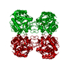
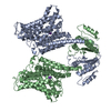
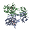
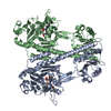
| Title | PURINE NUCLEOSIDE HYDROLASE WITH A TRANSITION STATE INHIBITOR | ||||||
 Components Components | INOSINE-URIDINE NUCLEOSIDE N-RIBOHYDROLASE | ||||||
 Keywords Keywords | HYDROLASE / PURINE NUCLEOSIDE HYDROLASE / INOSINE / URIDINE / IU-NH / PURINE NUCLEOSIDASE | ||||||
| Function / homology |  Function and homology information Function and homology informationinosine nucleosidase / uridine nucleosidase / inosine nucleosidase activity / uridine nucleosidase activity / purine nucleoside catabolic process / nucleotide metabolic process / metal ion binding / cytosol Similarity search - Function | ||||||
| Biological species |  Crithidia fasciculata (eukaryote) Crithidia fasciculata (eukaryote) | ||||||
| Method |  X-RAY DIFFRACTION / MOLECULAR REPLACEMENT / Resolution: 2.3 Å X-RAY DIFFRACTION / MOLECULAR REPLACEMENT / Resolution: 2.3 Å | ||||||
 Authors Authors | Degano, M. / Schramm, V.L. / Sacchettini, J.C. | ||||||
 Citation Citation | Journal: Biochemistry / Year: 1998 Title: Trypanosomal nucleoside hydrolase. A novel mechanism from the structure with a transition-state inhibitor. Authors: Degano, M. / Almo, S.C. / Sacchettini, J.C. / Schramm, V.L. #1: Journal: Biochemistry / Year: 1996Title: Three-Dimensional Structure of the Inosine-Uridine Nucleoside N-Ribohydrolase from Crithidia Fasciculata Authors: Degano, M. / Gopaul, D.N. / Scapin, G. / Schramm, V.L. / Sacchettini, J.C. | ||||||
| History |
|
- Structure visualization
| Structure viewer | Molecule: MolmilJmol/JSmol |
|---|
- Downloads & links
-Download
| PDBx/mmCIF format | 2mas.cif.gz | 250.3 KB | Display | PDBx/mmCIF format |
|---|---|---|---|---|
| PDB format | pdb2mas.ent.gz | 202.4 KB | Display | PDB format |
| PDBx/mmJSON format | 2mas.json.gz | Tree view | PDBx/mmJSON format | |
| Others |  Other downloads Other downloads |
-Validation report
| Arichive directory | https://data.pdbj.org/pub/pdb/validation_reports/ma/2masftp://data.pdbj.org/pub/pdb/validation_reports/ma/2mas | HTTPS FTP |
|---|
-Related structure data
| Related structure data | 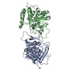 1masS S: Starting model for refinement |
|---|---|
| Similar structure data |


-Links
 PDBj
PDBj- Assembly
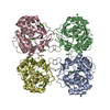
| Deposited unit | 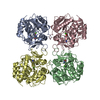
| ||||||||||||||||
|---|---|---|---|---|---|---|---|---|---|---|---|---|---|---|---|---|---|
| 1 |
| ||||||||||||||||
| Unit cell |
| ||||||||||||||||
| Noncrystallographic symmetry (NCS) | NCS oper:
|
-Components
| #1: Protein | Mass: 34206.480 Da / Num. of mol.: 4 / Mutation: P2A Source method: isolated from a genetically manipulated source Source: (gene. exp.) Crithidia fasciculata (eukaryote) / Gene: IU-NH FROM C. FASCICULATA / Plasmid: PET3D-IUNH / Gene (production host): IU-NH FROM C. FASCICULATA / Production host:  #2: Chemical | ChemComp-CA /   Mass: 40.078 Da / Num. of mol.: 4 / Source method: obtained synthetically / Formula: Ca Mass: 40.078 Da / Num. of mol.: 4 / Source method: obtained synthetically / Formula: Ca#3: Chemical | ChemComp-PIR /   Mass: 224.256 Da / Num. of mol.: 4 / Source method: obtained synthetically / Formula: C11H16N2O3 Mass: 224.256 Da / Num. of mol.: 4 / Source method: obtained synthetically / Formula: C11H16N2O3#4: Water | ChemComp-HOH / |  Mass: 18.015 Da / Num. of mol.: 158 / Source method: isolated from a natural source / Formula: H2O Mass: 18.015 Da / Num. of mol.: 158 / Source method: isolated from a natural source / Formula: H2O |
|---|
-Experimental details
-Experiment
| Experiment | Method: X-RAY DIFFRACTION / Number of used crystals: 1 |
|---|
- Sample preparation
| Crystal | Density Matthews: 3.58 Å3/Da / Density % sol: 65 % | ||||||||||||||||||||||||||||||||||||||||
|---|---|---|---|---|---|---|---|---|---|---|---|---|---|---|---|---|---|---|---|---|---|---|---|---|---|---|---|---|---|---|---|---|---|---|---|---|---|---|---|---|---|
| Crystal grow | pH: 7.5 / Details: 1.8 M AMMONIUM SULFATE, 3% ISOPROPANOL, pH 7.5 | ||||||||||||||||||||||||||||||||||||||||
| Crystal grow | *PLUS pH: 7 / Method: vapor diffusion, hanging drop | ||||||||||||||||||||||||||||||||||||||||
| Components of the solutions | *PLUS
|
-Data collection
| Diffraction | Mean temperature: 293 K |
|---|---|
| Diffraction source | Source: ROTATING ANODE / Type: RIGAKU / Wavelength: 1.5418 |
| Detector | Type: SIEMENS / Detector: AREA DETECTOR / Date: Oct 16, 1995 |
| Radiation | Monochromator: GRAPHITE(002) / Monochromatic (M) / Laue (L): M / Scattering type: x-ray |
| Radiation wavelength | Wavelength: 1.5418 Å / Relative weight: 1 |
| Reflection | Lowest resolution: 2.3 Å / Num. obs: 73166 / % possible obs: 80.8 % / Observed criterion σ(I): 0 / Redundancy: 4.5 % / Rmerge(I) obs: 0.082 / Net I/σ(I): 9.31 |
| Reflection shell | Resolution: 2.3→2.5 Å / Mean I/σ(I) obs: 2.2 / % possible all: 75.1 |
| Reflection | *PLUS Highest resolution: 2.3 Å |
| Reflection shell | *PLUS % possible obs: 75 % |
- Processing
| Software |
| ||||||||||||||||||||||||||||||||||||||||||||||||||||||||||||||||||||||||||||||||
|---|---|---|---|---|---|---|---|---|---|---|---|---|---|---|---|---|---|---|---|---|---|---|---|---|---|---|---|---|---|---|---|---|---|---|---|---|---|---|---|---|---|---|---|---|---|---|---|---|---|---|---|---|---|---|---|---|---|---|---|---|---|---|---|---|---|---|---|---|---|---|---|---|---|---|---|---|---|---|---|---|---|
| Refinement | Method to determine structure: MOLECULAR REPLACEMENT Starting model: UNLIGANDED IU-NH (PDB CODE 1MAS), WATERS AND IONS REMOVED Highest resolution: 2.3 Å / Data cutoff high absF: 10000000 / Data cutoff low absF: 0 / Cross valid method: THROUGHOUT / σ(F): 0
| ||||||||||||||||||||||||||||||||||||||||||||||||||||||||||||||||||||||||||||||||
| Displacement parameters | Biso mean: 29.1 Å2 | ||||||||||||||||||||||||||||||||||||||||||||||||||||||||||||||||||||||||||||||||
| Refine analyze | Luzzati coordinate error free: 0.35 Å | ||||||||||||||||||||||||||||||||||||||||||||||||||||||||||||||||||||||||||||||||
| Refinement step | Cycle: LAST / Highest resolution: 2.3 Å
| ||||||||||||||||||||||||||||||||||||||||||||||||||||||||||||||||||||||||||||||||
| Refine LS restraints |
| ||||||||||||||||||||||||||||||||||||||||||||||||||||||||||||||||||||||||||||||||
| Refine LS restraints NCS | Rms dev Biso : 1.5 Å2 / Rms dev position: 0.04 Å / Weight Biso : 3 / Weight position: 500 | ||||||||||||||||||||||||||||||||||||||||||||||||||||||||||||||||||||||||||||||||
| LS refinement shell | Resolution: 2.3→2.4 Å / Total num. of bins used: 8
| ||||||||||||||||||||||||||||||||||||||||||||||||||||||||||||||||||||||||||||||||
| Xplor file |
| ||||||||||||||||||||||||||||||||||||||||||||||||||||||||||||||||||||||||||||||||
| Software | *PLUS Name: X-PLOR / Version: 3.1 / Classification: refinement | ||||||||||||||||||||||||||||||||||||||||||||||||||||||||||||||||||||||||||||||||
| Refinement | *PLUS Lowest resolution: 25 Å | ||||||||||||||||||||||||||||||||||||||||||||||||||||||||||||||||||||||||||||||||
| Solvent computation | *PLUS | ||||||||||||||||||||||||||||||||||||||||||||||||||||||||||||||||||||||||||||||||
| Displacement parameters | *PLUS | ||||||||||||||||||||||||||||||||||||||||||||||||||||||||||||||||||||||||||||||||
| Refine LS restraints | *PLUS
| ||||||||||||||||||||||||||||||||||||||||||||||||||||||||||||||||||||||||||||||||
| LS refinement shell | *PLUS Rfactor obs: 0.323 |
