 Movie
Movie Controller

[English] 日本語
 Yorodumi
- PDB-1hnh: CRYSTAL STRUCTURE OF BETA-KETOACYL-ACP SYNTHASE III + DEGRADED FO... -
+ Open data

- Basic information
| Entry | Database: PDB / ID: 1hnh | ||||||
|---|---|---|---|---|---|---|---|
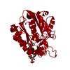
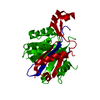
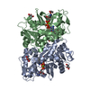
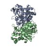
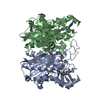
| Title | CRYSTAL STRUCTURE OF BETA-KETOACYL-ACP SYNTHASE III + DEGRADED FORM OF ACETYL-COA | ||||||
 Components Components | BETA-KETOACYL-ACYL CARRIER PROTEIN SYNTHASE III | ||||||
 Keywords Keywords | TRANSFERASE / FabH | ||||||
| Function / homology |  Function and homology information Function and homology informationbeta-ketoacyl-[acyl-carrier-protein] synthase III / beta-ketoacyl-acyl-carrier-protein synthase III activity / fatty acid elongation / 3-oxoacyl-[acyl-carrier-protein] synthase activity / fatty acid metabolic process / cytosol Similarity search - Function | ||||||
| Biological species |  | ||||||
| Method |  X-RAY DIFFRACTION / SYNCHROTRON / MAD / Resolution: 1.9 Å X-RAY DIFFRACTION / SYNCHROTRON / MAD / Resolution: 1.9 Å | ||||||
 Authors Authors | Qiu, X. / Janson, C.A. / Smith, W.W. / Head, M. / Lonsdale, J. / Konstantinidis, A.K. | ||||||
 Citation Citation | Journal: J.Mol.Biol. / Year: 2001 Title: Refined structures of beta-ketoacyl-acyl carrier protein synthase III. Authors: Qiu, X. / Janson, C.A. / Smith, W.W. / Head, M. / Lonsdale, J. / Konstantinidis, A.K. #1: Journal: J.Biol.Chem. / Year: 1999Title: Crystal Structure of Beta-Ketoacyl-Acyl Carrier Protein Synthase III. A Key Condensing Enzyme in Bacterial Fatty Acid Biosynthesis Authors: Qiu, X. / Janson, C.A. / Konstantinidis, A.K. / Nwagwu, S. / Silverman, C. / Smith, W.W. / Khandekar, S.K. / Lonsdale, J. / Abdel-Meguid, S.S. | ||||||
| History |
|
- Structure visualization
| Structure viewer | Molecule: MolmilJmol/JSmol |
|---|
- Downloads & links
-Download
| PDBx/mmCIF format | 1hnh.cif.gz | 77.8 KB | Display | PDBx/mmCIF format |
|---|---|---|---|---|
| PDB format | pdb1hnh.ent.gz | 58 KB | Display | PDB format |
| PDBx/mmJSON format | 1hnh.json.gz | Tree view | PDBx/mmJSON format | |
| Others |  Other downloads Other downloads |
-Validation report
| Arichive directory | https://data.pdbj.org/pub/pdb/validation_reports/hn/1hnhftp://data.pdbj.org/pub/pdb/validation_reports/hn/1hnh | HTTPS FTP |
|---|
-Related structure data


-Links
 PDBj
PDBj


- Assembly
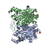
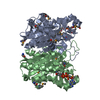
| Deposited unit | 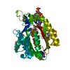
| ||||||||
|---|---|---|---|---|---|---|---|---|---|
| 1 | 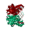
| ||||||||
| Unit cell |
| ||||||||
| Components on special symmetry positions |
|
-Components
| #1: Protein | Mass: 33964.223 Da / Num. of mol.: 1 Source method: isolated from a genetically manipulated source Source: (gene. exp.) References: UniProt: P0A6R0, beta-ketoacyl-[acyl-carrier-protein] synthase I |
|---|---|
| #2: Chemical | ChemComp-COA /   Mass: 767.534 Da / Num. of mol.: 1 / Source method: obtained synthetically / Formula: C21H36N7O16P3S Mass: 767.534 Da / Num. of mol.: 1 / Source method: obtained synthetically / Formula: C21H36N7O16P3S |
| #3: Water | ChemComp-HOH /  Mass: 18.015 Da / Num. of mol.: 236 / Source method: isolated from a natural source / Formula: H2O Mass: 18.015 Da / Num. of mol.: 236 / Source method: isolated from a natural source / Formula: H2O |
| Has protein modification | Y |
-Experimental details
-Experiment
| Experiment | Method: X-RAY DIFFRACTION / Number of used crystals: 1 |
|---|
- Sample preparation
| Crystal | Density Matthews: 1.99 Å3/Da / Density % sol: 38.04 % | ||||||||||||||||||||||||||||||||||||||||||||||||
|---|---|---|---|---|---|---|---|---|---|---|---|---|---|---|---|---|---|---|---|---|---|---|---|---|---|---|---|---|---|---|---|---|---|---|---|---|---|---|---|---|---|---|---|---|---|---|---|---|---|
| Crystal grow | Temperature: 298 K / Method: vapor diffusion, sitting drop / pH: 7 Details: PEK4000, pH 7, VAPOR DIFFUSION, SITTING DROP, temperature 298K | ||||||||||||||||||||||||||||||||||||||||||||||||
| Crystal grow | *PLUS pH: 7.4 / Method: vapor diffusion / Details: Janson, C.A., (2000) Acta Crystallogr.D, 56, 747. | ||||||||||||||||||||||||||||||||||||||||||||||||
| Components of the solutions | *PLUS
|
-Data collection
| Diffraction | Mean temperature: 100 K |
|---|---|
| Diffraction source | Source: SYNCHROTRON / Site: NSLS  / Beamline: X12C / Wavelength: 1 Å / Beamline: X12C / Wavelength: 1 Å |
| Detector | Type: CUSTOM-MADE / Detector: CCD / Date: Apr 1, 1999 |
| Radiation | Protocol: MAD / Monochromatic (M) / Laue (L): M / Scattering type: x-ray |
| Radiation wavelength | Wavelength: 1 Å / Relative weight: 1 |
| Reflection | Resolution: 1.9→20 Å / Num. obs: 203786 / % possible obs: 81 % / Observed criterion σ(F): 0 / Observed criterion σ(I): 0 / Redundancy: 6 % / Rmerge(I) obs: 0.083 |
| Reflection | *PLUS Num. obs: 33450 / Num. measured all: 203786 |
| Reflection shell | *PLUS % possible obs: 51.1 % / Rmerge(I) obs: 0.355 / Mean I/σ(I) obs: 4.9 |
- Processing
| Software |
| ||||||||||||||||
|---|---|---|---|---|---|---|---|---|---|---|---|---|---|---|---|---|---|
| Refinement | Method to determine structure: MAD / Resolution: 1.9→20 Å / σ(F): 0 / σ(I): 0 Details: THE ACETYL-CoA WAS DEGRADED TO COA. CYSTEINE 112 IS ACETYLATED AND IS LABELLED AS SCY.
| ||||||||||||||||
| Refinement step | Cycle: LAST / Resolution: 1.9→20 Å
| ||||||||||||||||
| Refine LS restraints |
| ||||||||||||||||
| Software | *PLUS Name: CNS / Classification: refinement | ||||||||||||||||
| Refinement | *PLUS Num. reflection all: 33450 / Num. reflection obs: 30884 | ||||||||||||||||
| Solvent computation | *PLUS | ||||||||||||||||
| Displacement parameters | *PLUS |
