 Movie
Movie Controller

[English] 日本語
 Yorodumi
- PDB-1hh7: REFINED CRYSTAL STRUCTURE OF CYTOCHROME C2 FROM RHODOPSEUDOMONAS ... -
+ Open data

- Basic information
| Entry | Database: PDB / ID: 1hh7 | |||||||||
|---|---|---|---|---|---|---|---|---|---|---|
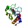
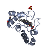
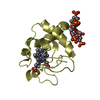
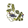
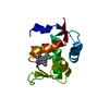
| Title | REFINED CRYSTAL STRUCTURE OF CYTOCHROME C2 FROM RHODOPSEUDOMONAS PALUSTRIS AT 1.4 ANGSTROM RESOLUTION | |||||||||
 Components Components | CYTOCHROME C2 | |||||||||
 Keywords Keywords | ELECTRON TRANSPORT / ELECTRON CARRIER / HEME PROTEIN | |||||||||
| Function / homology |  Function and homology information Function and homology informationphotosynthesis / electron transfer activity / heme binding / metal ion binding Similarity search - Function | |||||||||
| Biological species |  RHODOPSEUDOMONAS PALUSTRIS (phototrophic) RHODOPSEUDOMONAS PALUSTRIS (phototrophic) | |||||||||
| Method |  X-RAY DIFFRACTION / SYNCHROTRON / MOLECULAR REPLACEMENT / Resolution: 1.4 Å X-RAY DIFFRACTION / SYNCHROTRON / MOLECULAR REPLACEMENT / Resolution: 1.4 Å | |||||||||
 Authors Authors | Garau, G. / Geremia, S. | |||||||||
 Citation Citation | Journal: Acta Crystallogr.,Sect.D / Year: 2000 Title: Crystallization and Preliminary X-Ray Analysis of Two Ph-Dependent Forms of Cytochrome C2 from Rhodopseudomonas Palustris Authors: Garau, G. / Geremia, S. / Randaccio, L. / Vaccari, L. / Viezzoli, M.S. #1: Journal: J.Mol.Biol. / Year: 1995Title: Refined Crystal Structure of Ferrocytochrome C2 from Rhodopseudomonas Viridis at 1.6 A Resolution Authors: Sogabe, S. / Miki, K. #2: Journal: Nature / Year: 1979 Title: Cytochrome C2 Sequence Variation Among the Recognised Species of Purple Nonsulphur Photosynthetic Bacteria Authors: Ambler, R.P. / Daniel, M. / Hermoso, J. / Meyer, T.E. / Bartsch, R.G. / Kamen, M.D. | |||||||||
| History |
|
- Structure visualization
| Structure viewer | Molecule: MolmilJmol/JSmol |
|---|
- Downloads & links
-Download
| PDBx/mmCIF format | 1hh7.cif.gz | 59.2 KB | Display | PDBx/mmCIF format |
|---|---|---|---|---|
| PDB format | pdb1hh7.ent.gz | 39.9 KB | Display | PDB format |
| PDBx/mmJSON format | 1hh7.json.gz | Tree view | PDBx/mmJSON format | |
| Others |  Other downloads Other downloads |
-Validation report
| Summary document | 1hh7_validation.pdf.gz | 483.1 KB | Display | wwPDB validaton report |
|---|---|---|---|---|
| Full document | 1hh7_full_validation.pdf.gz | 485.7 KB | Display | |
| Data in XML | 1hh7_validation.xml.gz | 4.5 KB | Display | |
| Data in CIF | 1hh7_validation.cif.gz | 7.6 KB | Display | |
| Arichive directory | https://data.pdbj.org/pub/pdb/validation_reports/hh/1hh7ftp://data.pdbj.org/pub/pdb/validation_reports/hh/1hh7 | HTTPS FTP |
-Related structure data
| Related structure data |  2c2cS S: Starting model for refinement |
|---|---|
| Similar structure data |


-Links
 PDBj
PDBj


- Assembly
| Deposited unit | 
| ||||||||
|---|---|---|---|---|---|---|---|---|---|
| 1 |
| ||||||||
| Unit cell |
| ||||||||
| Components on special symmetry positions |
|
-Components
| #1: Protein | Mass: 12186.983 Da / Num. of mol.: 1 / Source method: isolated from a natural source / Source: (natural) RHODOPSEUDOMONAS PALUSTRIS (phototrophic) / Strain: 42 OL / References: UniProt: P00091 | ||||||||
|---|---|---|---|---|---|---|---|---|---|
| #2: Chemical | ChemComp-HEC /   Mass: 618.503 Da / Num. of mol.: 1 / Source method: obtained synthetically / Formula: C34H34FeN4O4 Mass: 618.503 Da / Num. of mol.: 1 / Source method: obtained synthetically / Formula: C34H34FeN4O4 | ||||||||
| #3: Chemical |   Mass: 96.063 Da / Num. of mol.: 2 / Source method: obtained synthetically / Formula: SO4 Mass: 96.063 Da / Num. of mol.: 2 / Source method: obtained synthetically / Formula: SO4#4: Chemical | ChemComp-NH3 / |   Mass: 17.031 Da / Num. of mol.: 1 / Source method: obtained synthetically / Formula: NH3 Mass: 17.031 Da / Num. of mol.: 1 / Source method: obtained synthetically / Formula: NH3#5: Water | ChemComp-HOH / |  Mass: 18.015 Da / Num. of mol.: 249 / Source method: isolated from a natural source / Formula: H2O Mass: 18.015 Da / Num. of mol.: 249 / Source method: isolated from a natural source / Formula: H2OHas protein modification | Y | Sequence details | PCA 1: GLN 1 HAS BEEN CYCLIZED TO PYRROLIDON | |
-Experimental details
-Experiment
| Experiment | Method: X-RAY DIFFRACTION / Number of used crystals: 1 |
|---|
- Sample preparation
| Crystal | Density Matthews: 3.18 Å3/Da / Density % sol: 61.3 % | ||||||||||||||||||||||||||||||
|---|---|---|---|---|---|---|---|---|---|---|---|---|---|---|---|---|---|---|---|---|---|---|---|---|---|---|---|---|---|---|---|
| Crystal grow | pH: 9 / Details: 60% AMMONIUM SULPHATE, 0.1 M TRIS PH 9.0 | ||||||||||||||||||||||||||||||
| Crystal grow | *PLUS Temperature: 291 K / pH: 6 / Method: vapor diffusion, hanging drop | ||||||||||||||||||||||||||||||
| Components of the solutions | *PLUS
|
-Data collection
| Diffraction | Mean temperature: 100 K |
|---|---|
| Diffraction source | Source: SYNCHROTRON / Site: ELETTRA  / Beamline: 5.2R / Wavelength: 1 / Beamline: 5.2R / Wavelength: 1 |
| Detector | Type: MARRESEARCH / Detector: IMAGE PLATE / Date: Apr 23, 1999 / Details: MIRRORS |
| Radiation | Monochromator: SI(111) / Protocol: SINGLE WAVELENGTH / Monochromatic (M) / Laue (L): M / Scattering type: x-ray |
| Radiation wavelength | Wavelength: 1 Å / Relative weight: 1 |
| Reflection | Resolution: 1.4→56 Å / Num. obs: 31246 / % possible obs: 99.8 % / Observed criterion σ(I): 0 / Redundancy: 12 % / Rmerge(I) obs: 0.096 / Net I/σ(I): 5.9 |
| Reflection shell | Resolution: 1.4→1.47 Å / Redundancy: 11.7 % / Rmerge(I) obs: 0.507 / Mean I/σ(I) obs: 5.9 / % possible all: 99.8 |
| Reflection | *PLUS Highest resolution: 1.4 Å / Lowest resolution: 56 Å / Observed criterion σ(I): 0 / Num. measured all: 394132 |
| Reflection shell | *PLUS % possible obs: 99.8 % |
- Processing
| Software |
| |||||||||||||||||||||||||||||||||
|---|---|---|---|---|---|---|---|---|---|---|---|---|---|---|---|---|---|---|---|---|---|---|---|---|---|---|---|---|---|---|---|---|---|---|
| Refinement | Method to determine structure: MOLECULAR REPLACEMENT Starting model: 2C2C Resolution: 1.4→56 Å / Num. parameters: 4681 / Num. restraintsaints: 3727 / Cross valid method: FREE R-VALUE / σ(F): 0 Details: ANISOTROPIC REFINEMENT FOR IRON AND ALL SULFUR ATOMS CHEMICALLY EQUIVALENT BONDS AND ANGLE DISTANCES IN HEME RESTRAINED TO BE EQUAL WITHOUT TARGET VALUES. NO GEOMETRIC OR ADP RESTRAINTS APPLIED TO IRON ATOM.
| |||||||||||||||||||||||||||||||||
| Refine analyze | Num. disordered residues: 1 | |||||||||||||||||||||||||||||||||
| Refinement step | Cycle: LAST / Resolution: 1.4→56 Å
| |||||||||||||||||||||||||||||||||
| Refine LS restraints |
| |||||||||||||||||||||||||||||||||
| Software | *PLUS Name: SHELX / Version: VERSION 97-1 / Classification: refinement | |||||||||||||||||||||||||||||||||
| Refine LS restraints | *PLUS
|
