 Movie
Movie Controller

[English] 日本語
 Yorodumi
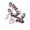
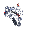
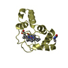
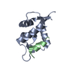
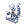
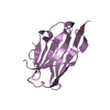
- PDB-1i8o: RHODOPSEUDOMONAS PALUSTRIS CYT C2 AMMONIA COMPLEX AT 1.15 ANGSTRO... -
+ Open data

- Basic information
| Entry | Database: PDB / ID: 1i8o | |||||||||
|---|---|---|---|---|---|---|---|---|---|---|
| Title | RHODOPSEUDOMONAS PALUSTRIS CYT C2 AMMONIA COMPLEX AT 1.15 ANGSTROM RESOLUTION | |||||||||
 Components Components | CYTOCHROME C2 | |||||||||
 Keywords Keywords | ELECTRON TRANSPORT / CYTOCHROME / HEME / AMMONIA / OXIDIZED | |||||||||
| Function / homology |  Function and homology information Function and homology informationphotosynthesis / electron transfer activity / heme binding / metal ion binding Similarity search - Function | |||||||||
| Biological species |  Rhodopseudomonas palustris (phototrophic) Rhodopseudomonas palustris (phototrophic) | |||||||||
| Method |  X-RAY DIFFRACTION / SYNCHROTRON / MOLECULAR REPLACEMENT / Resolution: 1.15 Å X-RAY DIFFRACTION / SYNCHROTRON / MOLECULAR REPLACEMENT / Resolution: 1.15 Å | |||||||||
 Authors Authors | Garau, G. / Geremia, S. | |||||||||
 Citation Citation | Journal: Protein Sci. / Year: 2002 Title: Cleavage of the iron-methionine bond in c-type cytochromes: crystal structure of oxidized and reduced cytochrome c(2) from Rhodopseudomonas palustris and its ammonia complex. Authors: Geremia, S. / Garau, G. / Vaccari, L. / Sgarra, R. / Viezzoli, M.S. / Calligaris, M. / Randaccio, L. #1: Journal: Acta Crystallogr.,Sect.D / Year: 2000Title: Crystallization and Preliminary X-Ray Analysis of Two Ph-Dependent Forms of Cytochrome C2 from Rhodopseudomonas Palustris Authors: Garau, G. / Geremia, S. / Randaccio, L. / Vaccari, L. / Viezzoli, M.S. #2: Journal: To be PublishedTitle: Crystal Structure of Oxidized and Reduced Cytochrome from Rhodopseudomonas Palustris: Relationship Between Hydrogen-Bonding Network of the Conservative Water Molecule and Reduction Potential Authors: Geremia, S. / Calligaris, M. / Garau, G. / Vaccari, L. / Viezzoli, M.S. / Randaccio, L. #3: Journal: Acta Crystallogr.,Sect.D / Year: 1994Title: Crystallization and X-Ray Structure Determination O Cytochrome C2 Fromrhodobacter Sphaeroides in Three Crystal Forms Authors: Axelrod, H. / Feher, G. / Allen, J.P. / Chirino, A.J. / Day, M.W. / Hsu, B.T. / Rees, D.C. #4: Journal: Nature / Year: 1979Title: Cytochrome C2 Sequence Variation Among the Recognised Species of Purple Nonsulphur Photosynthetic Bacteria Authors: Ambler, R.P. / Daniel, M. / Hermoso, J. / Meyer, T.E. / Bartsch, R.G. / Kamen, M.D. | |||||||||
| History |
| |||||||||
| Remark 999 | SEQUENCE PCA 1: GLN 1 HAS BEEN CYCLIZED TO PYRROLIDONE CARBOXYLIC ACID. THIS PROTEIN WAS EXPRESSED ...SEQUENCE PCA 1: GLN 1 HAS BEEN CYCLIZED TO PYRROLIDONE CARBOXYLIC ACID. THIS PROTEIN WAS EXPRESSED FROM A DIFFERENT STRAIN THAN THE DATABASE PROTEIN SWISSPROT ENTRY P00091 AND HAS SEVERAL MUTATIONS: G29A, I64V, N65P, N68A, AND D80E. |
- Structure visualization
| Structure viewer | Molecule: MolmilJmol/JSmol |
|---|
- Downloads & links
-Download
| PDBx/mmCIF format | 1i8o.cif.gz | 72 KB | Display | PDBx/mmCIF format |
|---|---|---|---|---|
| PDB format | pdb1i8o.ent.gz | 51.9 KB | Display | PDB format |
| PDBx/mmJSON format | 1i8o.json.gz | Tree view | PDBx/mmJSON format | |
| Others |  Other downloads Other downloads |
-Validation report
| Arichive directory | https://data.pdbj.org/pub/pdb/validation_reports/i8/1i8oftp://data.pdbj.org/pub/pdb/validation_reports/i8/1i8o | HTTPS FTP |
|---|
-Related structure data
| Related structure data |  1fj0C 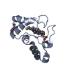 1i8pC  1hh7S C: citing same article ( S: Starting model for refinement |
|---|---|
| Similar structure data |


-Links
 PDBj
PDBj


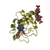
- Assembly
| Deposited unit | 
| ||||||||
|---|---|---|---|---|---|---|---|---|---|
| 1 |
| ||||||||
| 2 | 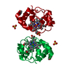
| ||||||||
| Unit cell |
| ||||||||
| Components on special symmetry positions |
|
-Components
| #1: Protein | Mass: 12186.983 Da / Num. of mol.: 1 / Source method: isolated from a natural source / Source: (natural) Rhodopseudomonas palustris (phototrophic) / Strain: 42 OL / References: UniProt: P00091 | ||||||||
|---|---|---|---|---|---|---|---|---|---|
| #2: Chemical |   Mass: 96.063 Da / Num. of mol.: 2 / Source method: obtained synthetically / Formula: SO4 Mass: 96.063 Da / Num. of mol.: 2 / Source method: obtained synthetically / Formula: SO4#3: Chemical | ChemComp-HEC / |   Mass: 618.503 Da / Num. of mol.: 1 / Source method: obtained synthetically / Formula: C34H34FeN4O4 Mass: 618.503 Da / Num. of mol.: 1 / Source method: obtained synthetically / Formula: C34H34FeN4O4#4: Chemical | ChemComp-NH3 / | 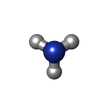  Mass: 17.031 Da / Num. of mol.: 1 / Source method: obtained synthetically / Formula: NH3 Mass: 17.031 Da / Num. of mol.: 1 / Source method: obtained synthetically / Formula: NH3#5: Water | ChemComp-HOH / |  Mass: 18.015 Da / Num. of mol.: 239 / Source method: isolated from a natural source / Formula: H2O Mass: 18.015 Da / Num. of mol.: 239 / Source method: isolated from a natural source / Formula: H2OHas protein modification | Y | |
-Experimental details
-Experiment
| Experiment | Method: X-RAY DIFFRACTION / Number of used crystals: 1 |
|---|
- Sample preparation
| Crystal | Density Matthews: 3.2 Å3/Da / Density % sol: 61 % | ||||||||||||||||||||||||||||||
|---|---|---|---|---|---|---|---|---|---|---|---|---|---|---|---|---|---|---|---|---|---|---|---|---|---|---|---|---|---|---|---|
| Crystal grow | Temperature: 293 K / Method: vapor diffusion, hanging drop / pH: 8.5 Details: 60% AMMONIUM SULPHATE, 0.1 M TRIS PH 8.5, VAPOR DIFFUSION, HANGING DROP, temperature 20K | ||||||||||||||||||||||||||||||
| Crystal grow | *PLUS pH: 6 | ||||||||||||||||||||||||||||||
| Components of the solutions | *PLUS
|
-Data collection
| Diffraction | Mean temperature: 100 K | |||||||||
|---|---|---|---|---|---|---|---|---|---|---|
| Diffraction source | Source: SYNCHROTRON / Site: ELETTRA  / Beamline: 5.2R / Wavelength: 1 / Wavelength: 1 Å / Beamline: 5.2R / Wavelength: 1 / Wavelength: 1 Å | |||||||||
| Detector | Type: MARRESEARCH / Detector: IMAGE PLATE / Date: Jul 8, 2000 / Details: MIRRORS | |||||||||
| Radiation | Monochromator: SI 111 / Protocol: SINGLE WAVELENGTH / Monochromatic (M) / Laue (L): M / Scattering type: x-ray | |||||||||
| Radiation wavelength |
| |||||||||
| Reflection | Resolution: 1.15→15.2 Å / Num. all: 59295 / Num. obs: 55707 / % possible obs: 99.1 % / Observed criterion σ(F): 2 / Observed criterion σ(I): 0 / Redundancy: 6.3 % / Biso Wilson estimate: 9.3 Å2 / Rmerge(I) obs: 0.054 / Net I/σ(I): 7.1 | |||||||||
| Reflection shell | Resolution: 1.15→1.21 Å / Redundancy: 5.9 % / Rmerge(I) obs: 0.412 / Mean I/σ(I) obs: 5.9 / % possible all: 99.1 | |||||||||
| Reflection | *PLUS Num. obs: 57362 / % possible obs: 99.3 % / Num. measured all: 364867 | |||||||||
| Reflection shell | *PLUS % possible obs: 96.7 % / Mean I/σ(I) obs: 4.1 |
- Processing
| Software |
| |||||||||||||||||||||||||
|---|---|---|---|---|---|---|---|---|---|---|---|---|---|---|---|---|---|---|---|---|---|---|---|---|---|---|
| Refinement | Method to determine structure: MOLECULAR REPLACEMENT Starting model: PDB ENTRY 1HH7 Resolution: 1.15→15.2 Å / Num. parameters: 10378 / Num. restraintsaints: 11837 / Cross valid method: FREE R / σ(F): 0 / σ(I): 2 / Stereochemistry target values: Engh & Huber
| |||||||||||||||||||||||||
| Refine analyze | Num. disordered residues: 1 | |||||||||||||||||||||||||
| Refinement step | Cycle: LAST / Resolution: 1.15→15.2 Å
| |||||||||||||||||||||||||
| Refine LS restraints |
| |||||||||||||||||||||||||
| Software | *PLUS Name: SHELX / Version: VERSION 97-1 / Classification: refinement | |||||||||||||||||||||||||
| Refinement | *PLUS Lowest resolution: 20 Å / % reflection Rfree: 5 % / Rfactor obs: 0.139 / Rfactor Rfree: 0.167 / Rfactor Rwork: 0.151 | |||||||||||||||||||||||||
| Solvent computation | *PLUS | |||||||||||||||||||||||||
| Displacement parameters | *PLUS | |||||||||||||||||||||||||
| Refine LS restraints | *PLUS
|
