 Movie
Movie Controller

[English] 日本語
 Yorodumi
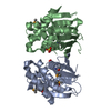
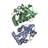
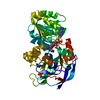
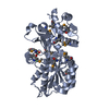
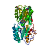
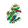
- PDB-1hfs: CRYSTAL STRUCTURE OF THE CATALYTIC DOMAIN OF HUMAN FIBROBLAST STR... -
+ Open data

- Basic information
| Entry | Database: PDB / ID: 1hfs | |||||||||
|---|---|---|---|---|---|---|---|---|---|---|
| Title | CRYSTAL STRUCTURE OF THE CATALYTIC DOMAIN OF HUMAN FIBROBLAST STROMELYSIN-1 INHIBITED WITH THE N-CARBOXY-ALKYL INHIBITOR L-764,004 | |||||||||
 Components Components | STROMELYSIN-1 | |||||||||
 Keywords Keywords | HYDROLASE / METALLOPROTEASE / MATRIX METALLOPROTEASE-3 / PROTEOGLYCANASE | |||||||||
| Function / homology |  Function and homology information Function and homology informationstromelysin 1 / cellular response to UV-A / regulation of neuroinflammatory response / Assembly of collagen fibrils and other multimeric structures / Activation of Matrix Metalloproteinases / response to amyloid-beta / Collagen degradation / negative regulation of reactive oxygen species metabolic process / collagen catabolic process / extracellular matrix disassembly ...stromelysin 1 / cellular response to UV-A / regulation of neuroinflammatory response / Assembly of collagen fibrils and other multimeric structures / Activation of Matrix Metalloproteinases / response to amyloid-beta / Collagen degradation / negative regulation of reactive oxygen species metabolic process / collagen catabolic process / extracellular matrix disassembly / Degradation of the extracellular matrix / extracellular matrix organization / cellular response to nitric oxide / protein catabolic process / EGFR Transactivation by Gastrin / negative regulation of phosphatidylinositol 3-kinase/protein kinase B signal transduction / cellular response to reactive oxygen species / positive regulation of protein-containing complex assembly / metalloendopeptidase activity / metallopeptidase activity / peptidase activity / cellular response to lipopolysaccharide / extracellular matrix / Interleukin-4 and Interleukin-13 signaling / endopeptidase activity / Extra-nuclear estrogen signaling / serine-type endopeptidase activity / innate immune response / mitochondrion / proteolysis / : / extracellular region / zinc ion binding / nucleus / cytosol Similarity search - Function | |||||||||
| Biological species |  Homo sapiens (human) Homo sapiens (human) | |||||||||
| Method |  X-RAY DIFFRACTION / Resolution: 1.7 Å X-RAY DIFFRACTION / Resolution: 1.7 Å | |||||||||
 Authors Authors | Becker, J.W. | |||||||||
 Citation Citation | Journal: J.Med.Chem. / Year: 1997 Title: Inhibition of stromelysin-1 (MMP-3) by P1'-biphenylylethyl carboxyalkyl dipeptides. Authors: Esser, C.K. / Bugianesi, R.L. / Caldwell, C.G. / Chapman, K.T. / Durette, P.L. / Girotra, N.N. / Kopka, I.E. / Lanza, T.J. / Levorse, D.A. / MacCoss, M. / Owens, K.A. / Ponpipom, M.M. / ...Authors: Esser, C.K. / Bugianesi, R.L. / Caldwell, C.G. / Chapman, K.T. / Durette, P.L. / Girotra, N.N. / Kopka, I.E. / Lanza, T.J. / Levorse, D.A. / MacCoss, M. / Owens, K.A. / Ponpipom, M.M. / Simeone, J.P. / Harrison, R.K. / Niedzwiecki, L. / Becker, J.W. / Marcy, A.I. / Axel, M.G. / Christen, A.J. / McDonnell, J. / Moore, V.L. / Olszewski, J.M. / Saphos, C. / Visco, D.M. / Shen, F. / Colletti, A. / Krieter, P.A. / Hagmann, W.K. #1: Journal: J.Biomol.NMR / Year: 1996Title: Comparison of the Structure of Human Recombinant Short Form Stromelysin by Multidimensional Heteronuclear NMR and X-Ray Crystallography Authors: Gooley, P.R. / O'Connell, J.F. / Marcy, A.I. / Cuca, G.C. / Axel, M.G. / Caldwell, C.G. / Hagmann, W.K. / Becker, J.W. #2: Journal: Protein Sci. / Year: 1995Title: Stromelysin-1: Three-Dimensional Structure of the Inhibited Catalytic Domain and of the C-Truncated Proenzyme Authors: Becker, J.W. / Marcy, A.I. / Rokosz, L.L. / Axel, M.G. / Burbaum, J.J. / Fitzgerald, P.M. / Cameron, P.M. / Esser, C.K. / Hagmann, W.K. / Hermes, J.D. / Springer, J.P. #3: Journal: Nat.Struct.Biol. / Year: 1994Title: The NMR Structure of the Inhibited Catalytic Domain of Human Stromelysin-1 Authors: Gooley, P.R. / O'Connell, J.F. / Marcy, A.I. / Cuca, G.C. / Salowe, S.P. / Bush, B.L. / Hermes, J.D. / Esser, C.K. / Hagmann, W.K. / Springer, J.P. / Johnson, B.A. #4: Journal: J.Med.Chem. / Year: 1993Title: Inhibition of Matrix Metalloproteinases by N-Carboxyalkyl Peptides Authors: Chapman, K.T. / Kopka, I.E. / Durette, P.L. / Esser, C.K. / Lanza, T.J. / Izquierdo-Martin, M. / Niedzwiecki, L. / Chang, B. / Harrison, R.K. / Kuo, D.W. / Lin, T.Y. / Stein, R.L. / Hagmann, W.K. #5: Journal: Biochemistry / Year: 1991Title: Human Fibroblast Stromelysin Catalytic Domain: Expression, Purification, and Characterization of a C-Terminally Truncated Form Authors: Marcy, A.I. / Eiberger, L.L. / Harrison, R. / Chan, H.K. / Hutchinson, N.I. / Hagmann, W.K. / Cameron, P.M. / Boulton, D.A. / Hermes, J.D. | |||||||||
| History |
|
- Structure visualization
| Structure viewer | Molecule: MolmilJmol/JSmol |
|---|
- Downloads & links
-Download
| PDBx/mmCIF format | 1hfs.cif.gz | 52.5 KB | Display | PDBx/mmCIF format |
|---|---|---|---|---|
| PDB format | pdb1hfs.ent.gz | 36.1 KB | Display | PDB format |
| PDBx/mmJSON format | 1hfs.json.gz | Tree view | PDBx/mmJSON format | |
| Others |  Other downloads Other downloads |
-Validation report
| Arichive directory | https://data.pdbj.org/pub/pdb/validation_reports/hf/1hfsftp://data.pdbj.org/pub/pdb/validation_reports/hf/1hfs | HTTPS FTP |
|---|
-Related structure data
| Similar structure data |
|---|


-Links
 PDBj
PDBj


- Assembly
| Deposited unit | 
| |||||||||
|---|---|---|---|---|---|---|---|---|---|---|
| 1 | 
| |||||||||
| Unit cell |
| |||||||||
| Components on special symmetry positions |
|
-Components
| #1: Protein | Mass: 17945.922 Da / Num. of mol.: 1 Source method: isolated from a genetically manipulated source Source: (gene. exp.) Homo sapiens (human) / Production host:  | ||||||
|---|---|---|---|---|---|---|---|
| #2: Chemical |   Mass: 65.409 Da / Num. of mol.: 2 / Source method: obtained synthetically / Formula: Zn Mass: 65.409 Da / Num. of mol.: 2 / Source method: obtained synthetically / Formula: Zn#3: Chemical |   Mass: 40.078 Da / Num. of mol.: 3 / Source method: obtained synthetically / Formula: Ca Mass: 40.078 Da / Num. of mol.: 3 / Source method: obtained synthetically / Formula: Ca#4: Chemical | ChemComp-L04 / | 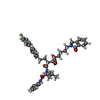  Mass: 705.857 Da / Num. of mol.: 1 / Source method: obtained synthetically / Formula: C43H48FN3O5 Mass: 705.857 Da / Num. of mol.: 1 / Source method: obtained synthetically / Formula: C43H48FN3O5#5: Water | ChemComp-HOH / |  Mass: 18.015 Da / Num. of mol.: 143 / Source method: isolated from a natural source / Formula: H2O Mass: 18.015 Da / Num. of mol.: 143 / Source method: isolated from a natural source / Formula: H2O |
-Experimental details
-Experiment
| Experiment | Method: X-RAY DIFFRACTION |
|---|
- Sample preparation
| Crystal | Density Matthews: 2.76 Å3/Da / Density % sol: 53 % | ||||||||||||||||||||||||||||||||||||||||||||||||||||||||||||
|---|---|---|---|---|---|---|---|---|---|---|---|---|---|---|---|---|---|---|---|---|---|---|---|---|---|---|---|---|---|---|---|---|---|---|---|---|---|---|---|---|---|---|---|---|---|---|---|---|---|---|---|---|---|---|---|---|---|---|---|---|---|
| Crystal grow | *PLUS pH: 6.5 / Method: vapor diffusion, hanging drop | ||||||||||||||||||||||||||||||||||||||||||||||||||||||||||||
| Components of the solutions | *PLUS
|
-Data collection
| Diffraction source | Wavelength: 1.5418 |
|---|---|
| Detector | Type: RIGAKU / Detector: IMAGE PLATE / Date: Aug 29, 1994 |
| Radiation | Monochromatic (M) / Laue (L): M / Scattering type: x-ray |
| Radiation wavelength | Wavelength: 1.5418 Å / Relative weight: 1 |
| Reflection | Num. obs: 21587 / % possible obs: 97 % / Observed criterion σ(I): 0 / Redundancy: 3.17 % / Biso Wilson estimate: 19.5 Å2 / Rmerge(I) obs: 0.0338 |
- Processing
| Software |
| ||||||||||||||||||||||||||||||||||||||||||||||||||||||||||||||||||||||||||||||||
|---|---|---|---|---|---|---|---|---|---|---|---|---|---|---|---|---|---|---|---|---|---|---|---|---|---|---|---|---|---|---|---|---|---|---|---|---|---|---|---|---|---|---|---|---|---|---|---|---|---|---|---|---|---|---|---|---|---|---|---|---|---|---|---|---|---|---|---|---|---|---|---|---|---|---|---|---|---|---|---|---|---|
| Refinement | Resolution: 1.7→20 Å / Rfactor Rfree error: 0.007 / Data cutoff high absF: 10000000 / Data cutoff low absF: 0.001 / Isotropic thermal model: RESTRAINED / Cross valid method: THROUGHOUT / σ(F): 2 / Details: BULK SOLVENT MODEL USED
| ||||||||||||||||||||||||||||||||||||||||||||||||||||||||||||||||||||||||||||||||
| Displacement parameters | Biso mean: 21.2 Å2 | ||||||||||||||||||||||||||||||||||||||||||||||||||||||||||||||||||||||||||||||||
| Refine analyze |
| ||||||||||||||||||||||||||||||||||||||||||||||||||||||||||||||||||||||||||||||||
| Refinement step | Cycle: LAST / Resolution: 1.7→20 Å
| ||||||||||||||||||||||||||||||||||||||||||||||||||||||||||||||||||||||||||||||||
| Refine LS restraints |
| ||||||||||||||||||||||||||||||||||||||||||||||||||||||||||||||||||||||||||||||||
| LS refinement shell | Resolution: 1.7→1.73 Å / Rfactor Rfree error: 0.049 / Total num. of bins used: 20
| ||||||||||||||||||||||||||||||||||||||||||||||||||||||||||||||||||||||||||||||||
| Xplor file |
| ||||||||||||||||||||||||||||||||||||||||||||||||||||||||||||||||||||||||||||||||
| Software | *PLUS Name: X-PLOR / Version: 3.1 / Classification: refinement | ||||||||||||||||||||||||||||||||||||||||||||||||||||||||||||||||||||||||||||||||
| Refine LS restraints | *PLUS
|
