 Movie
Movie Controller

[English] 日本語
 Yorodumi
- PDB-3on3: The crystal structure of keto/oxoacid ferredoxin oxidoreductase, ... -
+ Open data

- Basic information
| Entry | Database: PDB / ID: 3on3 | ||||||
|---|---|---|---|---|---|---|---|
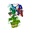
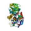
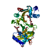
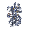
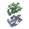
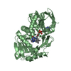
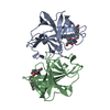
| Title | The crystal structure of keto/oxoacid ferredoxin oxidoreductase, gamma subunit from Geobacter sulfurreducens PCA | ||||||
 Components Components | Keto/oxoacid ferredoxin oxidoreductase, gamma subunit | ||||||
 Keywords Keywords | OXIDOREDUCTASE / structural genomics / PSI-2 / protein structure initiative / midwest center for structural genomics / MCSG | ||||||
| Function / homology |  Function and homology information Function and homology information | ||||||
| Biological species |  Geobacter sulfurreducens (bacteria) Geobacter sulfurreducens (bacteria) | ||||||
| Method |  X-RAY DIFFRACTION / SYNCHROTRON / SAD / Resolution: 2.193 Å X-RAY DIFFRACTION / SYNCHROTRON / SAD / Resolution: 2.193 Å | ||||||
 Authors Authors | Tan, K. / Zhang, R. / Hatzos, C. / Buck, K. / Joachimiak, A. / Midwest Center for Structural Genomics (MCSG) | ||||||
 Citation Citation | Journal: To be Published Title: The crystal structure of keto/oxoacid ferredoxin oxidoreductase, gamma subunit from Geobacter sulfurreducens PCA Authors: Tan, K. / Zhang, R. / Hatzos, C. / Buck, K. / Joachimiak, A. | ||||||
| History |
|
- Structure visualization
| Structure viewer | Molecule: MolmilJmol/JSmol |
|---|
- Downloads & links
-Download
| PDBx/mmCIF format | 3on3.cif.gz | 139 KB | Display | PDBx/mmCIF format |
|---|---|---|---|---|
| PDB format | pdb3on3.ent.gz | 111 KB | Display | PDB format |
| PDBx/mmJSON format | 3on3.json.gz | Tree view | PDBx/mmJSON format | |
| Others |  Other downloads Other downloads |
-Validation report
| Arichive directory | https://data.pdbj.org/pub/pdb/validation_reports/on/3on3ftp://data.pdbj.org/pub/pdb/validation_reports/on/3on3 | HTTPS FTP |
|---|
-Related structure data
| Similar structure data | |
|---|---|
| Other databases |


-Links
 PDBj
PDBj- Assembly
| Deposited unit | 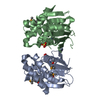
| ||||||||
|---|---|---|---|---|---|---|---|---|---|
| 1 |
| ||||||||
| 2 | 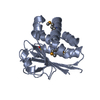
| ||||||||
| 3 | 
| ||||||||
| Unit cell |
| ||||||||
| Details | Experimentally unknown. It is predicted that the chains A and B are monomeric. |
-Components
| #1: Protein | Mass: 19272.404 Da / Num. of mol.: 2 Source method: isolated from a genetically manipulated source Source: (gene. exp.) Geobacter sulfurreducens (bacteria) / Strain: PCA / Gene: GSU1470 / Plasmid: pMCSG7 / Production host: #2: Chemical |   Mass: 96.063 Da / Num. of mol.: 2 / Source method: obtained synthetically / Formula: SO4 Mass: 96.063 Da / Num. of mol.: 2 / Source method: obtained synthetically / Formula: SO4#3: Water | ChemComp-HOH / |  Mass: 18.015 Da / Num. of mol.: 46 / Source method: isolated from a natural source / Formula: H2O Mass: 18.015 Da / Num. of mol.: 46 / Source method: isolated from a natural source / Formula: H2OHas protein modification | Y | |
|---|
-Experimental details
-Experiment
| Experiment | Method: X-RAY DIFFRACTION / Number of used crystals: 1 |
|---|
- Sample preparation
| Crystal | Density Matthews: 2.32 Å3/Da / Density % sol: 46.92 % |
|---|---|
| Crystal grow | Temperature: 289 K / Method: vapor diffusion, sitting drop / pH: 6.5 Details: 0.2M sodium chloride, 0.1M Sodium cacodylate, 2M Ammonium sulfate, pH 6.5, VAPOR DIFFUSION, SITTING DROP, temperature 289K |
-Data collection
| Diffraction | Mean temperature: 100 K |
|---|---|
| Diffraction source | Source: SYNCHROTRON / Site: APS  / Beamline: 19-ID / Wavelength: 0.97926 Å / Beamline: 19-ID / Wavelength: 0.97926 Å |
| Detector | Type: ADSC QUANTUM 315r / Detector: CCD / Date: Jun 7, 2010 / Details: mirror |
| Radiation | Monochromator: Si 111 crystal / Protocol: SINGLE WAVELENGTH / Monochromatic (M) / Laue (L): M / Scattering type: x-ray |
| Radiation wavelength | Wavelength: 0.97926 Å / Relative weight: 1 |
| Reflection | Resolution: 2.193→40 Å / Num. all: 18254 / Num. obs: 18254 / % possible obs: 98 % / Observed criterion σ(F): 0 / Observed criterion σ(I): 0 / Redundancy: 9.2 % / Rmerge(I) obs: 0.155 / Net I/σ(I): 20.6 |
| Reflection shell | Resolution: 2.2→2.24 Å / Redundancy: 7.8 % / Rmerge(I) obs: 0.827 / Mean I/σ(I) obs: 1.5 / Num. unique all: 796 / % possible all: 85.7 |
- Processing
| Software |
| |||||||||||||||||||||||||||||||||||||||||||||||||||||||||||||||||||||||||||
|---|---|---|---|---|---|---|---|---|---|---|---|---|---|---|---|---|---|---|---|---|---|---|---|---|---|---|---|---|---|---|---|---|---|---|---|---|---|---|---|---|---|---|---|---|---|---|---|---|---|---|---|---|---|---|---|---|---|---|---|---|---|---|---|---|---|---|---|---|---|---|---|---|---|---|---|---|
| Refinement | Method to determine structure: SAD / Resolution: 2.193→39.548 Å / SU ML: 0.32 / σ(F): 0.02 / σ(I): 0 / Stereochemistry target values: ML
| |||||||||||||||||||||||||||||||||||||||||||||||||||||||||||||||||||||||||||
| Solvent computation | Shrinkage radii: 0.9 Å / VDW probe radii: 1.11 Å / Solvent model: FLAT BULK SOLVENT MODEL / Bsol: 62.117 Å2 / ksol: 0.351 e/Å3 | |||||||||||||||||||||||||||||||||||||||||||||||||||||||||||||||||||||||||||
| Displacement parameters |
| |||||||||||||||||||||||||||||||||||||||||||||||||||||||||||||||||||||||||||
| Refinement step | Cycle: LAST / Resolution: 2.193→39.548 Å
| |||||||||||||||||||||||||||||||||||||||||||||||||||||||||||||||||||||||||||
| Refine LS restraints |
| |||||||||||||||||||||||||||||||||||||||||||||||||||||||||||||||||||||||||||
| LS refinement shell |
| |||||||||||||||||||||||||||||||||||||||||||||||||||||||||||||||||||||||||||
| Refinement TLS params. | Method: refined / Refine-ID: X-RAY DIFFRACTION
| |||||||||||||||||||||||||||||||||||||||||||||||||||||||||||||||||||||||||||
| Refinement TLS group |
|
