 Movie
Movie Controller


+ Open data

- Basic information
| Entry | Database: PDB / ID: 1hbo | ||||||
|---|---|---|---|---|---|---|---|
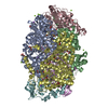
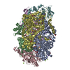
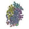
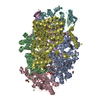
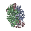
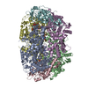
| Title | METHYL-COENZYME M REDUCTASE MCR-RED1-SILENT | ||||||
 Components Components | (METHYL-COENZYME M REDUCTASE I ...) x 3 | ||||||
 Keywords Keywords | METHANOGENESIS / BIOLOGICAL METHANOGENESIS / NI-ENZYME / OXIDOREDUCTASE | ||||||
| Function / homology |  Function and homology information Function and homology informationcoenzyme-B sulfoethylthiotransferase / coenzyme-B sulfoethylthiotransferase activity / methanogenesis / metal ion binding / cytoplasm Similarity search - Function | ||||||
| Biological species |   METHANOTHERMOBACTER THERMAUTOTROPHICUS (archaea) METHANOTHERMOBACTER THERMAUTOTROPHICUS (archaea) | ||||||
| Method |  X-RAY DIFFRACTION / MOLECULAR REPLACEMENT / Resolution: 1.78 Å X-RAY DIFFRACTION / MOLECULAR REPLACEMENT / Resolution: 1.78 Å | ||||||
 Authors Authors | Grabarse, W. | ||||||
 Citation Citation | Journal: J.Mol.Biol. / Year: 2001 Title: On the Mechanism of Biological Methane Formation: Structural Evidence for Conformational Changes in Methyl-Coenzyme M Reductase Upon Substrate Binding Authors: Grabarse, W. / Mahlert, F. / Duin, E.C. / Goubeaud, M. / Shima, S. / Thauer, R.K. / Lamzin, V. / Ermler, U. | ||||||
| History |
|
- Structure visualization
| Structure viewer | Molecule: MolmilJmol/JSmol |
|---|
- Downloads & links
-Download
| PDBx/mmCIF format | 1hbo.cif.gz | 549.3 KB | Display | PDBx/mmCIF format |
|---|---|---|---|---|
| PDB format | pdb1hbo.ent.gz | 439.7 KB | Display | PDB format |
| PDBx/mmJSON format | 1hbo.json.gz | Tree view | PDBx/mmJSON format | |
| Others |  Other downloads Other downloads |
-Validation report
| Arichive directory | https://data.pdbj.org/pub/pdb/validation_reports/hb/1hboftp://data.pdbj.org/pub/pdb/validation_reports/hb/1hbo | HTTPS FTP |
|---|
-Related structure data
| Related structure data |  1hbmC  1hbnC  1hbuC  1mroS C: citing same article ( S: Starting model for refinement |
|---|---|
| Similar structure data |


-Links
 PDBj
PDBj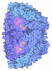
- Assembly
| Deposited unit | 
| ||||||||
|---|---|---|---|---|---|---|---|---|---|
| 1 |
| ||||||||
| Unit cell |
| ||||||||
| Noncrystallographic symmetry (NCS) | NCS oper: (Code: given Matrix: (0.6268, -0.7779, 0.0458), Vector: |
-Components
-METHYL-COENZYME M REDUCTASE I ... , 3 types, 6 molecules ADBECF
| #1: Protein | Mass: 60508.469 Da / Num. of mol.: 2 / Source method: isolated from a natural source Source: (natural) METHANOTHERMOBACTER THERMAUTOTROPHICUS (archaea)Cellular location: CYTOPLASM / Strain: MARBURG / References: UniProt: P11558 #2: Protein | Mass: 47148.477 Da / Num. of mol.: 2 / Source method: isolated from a natural source Source: (natural) METHANOTHERMOBACTER THERMAUTOTROPHICUS (archaea)Cellular location: CYTOPLASM / Strain: MARBURG / References: UniProt: P11560 #3: Protein | Mass: 28666.039 Da / Num. of mol.: 2 / Source method: isolated from a natural source / Details: MCR-OX-SILENT STATE, ENZYME-SUBSTRATE COMPLEX Source: (natural) METHANOTHERMOBACTER THERMAUTOTROPHICUS (archaea)Cellular location: CYTOPLASM / Strain: MARBURG / References: UniProt: P11562 |
|---|
-Non-polymers , 9 types, 2126 molecules 


| #4: Chemical |  Mass: 906.580 Da / Num. of mol.: 2 / Source method: obtained synthetically / Formula: C42H51N6NiO13 Mass: 906.580 Da / Num. of mol.: 2 / Source method: obtained synthetically / Formula: C42H51N6NiO13#5: Chemical |  Mass: 343.334 Da / Num. of mol.: 2 / Source method: obtained synthetically / Formula: C11H22NO7PS Mass: 343.334 Da / Num. of mol.: 2 / Source method: obtained synthetically / Formula: C11H22NO7PS#6: Chemical |  Mass: 142.197 Da / Num. of mol.: 2 / Source method: obtained synthetically / Formula: C2H6O3S2 Mass: 142.197 Da / Num. of mol.: 2 / Source method: obtained synthetically / Formula: C2H6O3S2#7: Chemical | ChemComp-GOL /  Mass: 92.094 Da / Num. of mol.: 9 / Source method: obtained synthetically / Formula: C3H8O3 Mass: 92.094 Da / Num. of mol.: 9 / Source method: obtained synthetically / Formula: C3H8O3#8: Chemical | ChemComp-ZN / |  Mass: 65.409 Da / Num. of mol.: 1 / Source method: obtained synthetically / Formula: Zn Mass: 65.409 Da / Num. of mol.: 1 / Source method: obtained synthetically / Formula: Zn#9: Chemical | ChemComp-NA /  Mass: 22.990 Da / Num. of mol.: 8 / Source method: obtained synthetically / Formula: Na Mass: 22.990 Da / Num. of mol.: 8 / Source method: obtained synthetically / Formula: Na#10: Chemical | ChemComp-MG /  Mass: 24.305 Da / Num. of mol.: 4 / Source method: obtained synthetically / Formula: Mg Mass: 24.305 Da / Num. of mol.: 4 / Source method: obtained synthetically / Formula: Mg#11: Chemical |  Mass: 35.453 Da / Num. of mol.: 2 / Source method: obtained synthetically / Formula: Cl Mass: 35.453 Da / Num. of mol.: 2 / Source method: obtained synthetically / Formula: Cl#12: Water | ChemComp-HOH / | Mass: 18.015 Da / Num. of mol.: 2096 / Source method: isolated from a natural source / Formula: H2O |
|---|
-Details
| Sequence details | REFERENCE: 1 RESIDUE IS LACKING IN THE C-TERMINUS AND THE N-TERMINUS, C AND N-TERMINUS ARE VISIBLE ...REFERENCE: 1 RESIDUE IS LACKING IN THE C-TERMINUS AND THE N-TERMINUS, C AND N-TERMINUS ARE VISIBLE IN THE CRYSTAL STRUCTURE. MET 1 WAS CLEARLY SHOWN NOT TO BE PRESENT. |
|---|
-Experimental details
-Experiment
| Experiment | Method: X-RAY DIFFRACTION / Number of used crystals: 1 |
|---|
- Sample preparation
| Crystal | Density Matthews: 3.9 Å3/Da / Density % sol: 31 % | ||||||||||||||||||||||||||||||||||||||||||||||||
|---|---|---|---|---|---|---|---|---|---|---|---|---|---|---|---|---|---|---|---|---|---|---|---|---|---|---|---|---|---|---|---|---|---|---|---|---|---|---|---|---|---|---|---|---|---|---|---|---|---|
| Crystal grow | pH: 7.5 Details: 25% PEG 400, 0.2 M NACL 20 MG/ML FINAL PROTEIN CONC 0.2 M MGCL, 0.1 M HEPES PH 7.5 | ||||||||||||||||||||||||||||||||||||||||||||||||
| Crystal grow | *PLUS Temperature: 4 ℃ / Method: vapor diffusion, hanging drop | ||||||||||||||||||||||||||||||||||||||||||||||||
| Components of the solutions | *PLUS
|
-Data collection
| Diffraction | Mean temperature: 100 K |
|---|---|
| Diffraction source | Source: ROTATING ANODE / Type: RIGAKU RU200 / Wavelength: 1.54 |
| Detector | Type: MAR scanner 345 mm plate / Detector: IMAGE PLATE / Date: Aug 15, 1998 |
| Radiation | Protocol: SINGLE WAVELENGTH / Monochromatic (M) / Laue (L): M / Scattering type: x-ray |
| Radiation wavelength | Wavelength: 1.54 Å / Relative weight: 1 |
| Reflection | Resolution: 1.78→10 Å / Num. obs: 211492 / % possible obs: 96.5 % / Redundancy: 3 % / Rmerge(I) obs: 0.058 / Rsym value: 0.06 / Net I/σ(I): 18.8 |
| Reflection shell | Resolution: 1.78→1.8 Å / Redundancy: 3.2 % / Rmerge(I) obs: 0.214 / Mean I/σ(I) obs: 5.36 / Rsym value: 0.19 / % possible all: 89 |
| Reflection | *PLUS Lowest resolution: 10 Å / Redundancy: 3.2 % |
| Reflection shell | *PLUS % possible obs: 89.2 % / Redundancy: 2.94 % |
- Processing
| Software |
| |||||||||||||||||||||||||||||||||||||||||||||||||||||||||||||||
|---|---|---|---|---|---|---|---|---|---|---|---|---|---|---|---|---|---|---|---|---|---|---|---|---|---|---|---|---|---|---|---|---|---|---|---|---|---|---|---|---|---|---|---|---|---|---|---|---|---|---|---|---|---|---|---|---|---|---|---|---|---|---|---|---|
| Refinement | Method to determine structure: MOLECULAR REPLACEMENT Starting model: 1MRO Resolution: 1.78→10 Å / Cross valid method: THROUGHOUT / σ(F): 0
| |||||||||||||||||||||||||||||||||||||||||||||||||||||||||||||||
| Refinement step | Cycle: LAST / Resolution: 1.78→10 Å
| |||||||||||||||||||||||||||||||||||||||||||||||||||||||||||||||
| Refine LS restraints |
| |||||||||||||||||||||||||||||||||||||||||||||||||||||||||||||||
| Software | *PLUS Name: REFMAC / Classification: refinement | |||||||||||||||||||||||||||||||||||||||||||||||||||||||||||||||
| Refinement | *PLUS Rfactor obs: 0.177 | |||||||||||||||||||||||||||||||||||||||||||||||||||||||||||||||
| Solvent computation | *PLUS | |||||||||||||||||||||||||||||||||||||||||||||||||||||||||||||||
| Displacement parameters | *PLUS |
