 Movie
Movie Controller

[English] 日本語
 Yorodumi
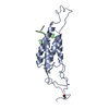
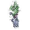
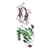
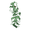
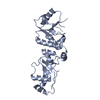
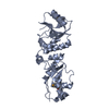
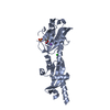
- PDB-1h6e: MU2 ADAPTIN SUBUNIT (AP50) OF AP2 ADAPTOR (SECOND DOMAIN), COMPLE... -
+ Open data

- Basic information
| Entry | Database: PDB / ID: 1h6e | |||||||||
|---|---|---|---|---|---|---|---|---|---|---|
| Title | MU2 ADAPTIN SUBUNIT (AP50) OF AP2 ADAPTOR (SECOND DOMAIN), COMPLEXED WITH CTLA-4 INTERNALIZATION PEPTIDE TTGVYVKMPPT | |||||||||
 Components Components |
| |||||||||
 Keywords Keywords | ENDOCYTOSIS/EXOCYTOSIS / ENDOCYTOSIS / ADAPTOR / PEPTIDE COMPLEX / PHOSPHORYLATION / ENDOCYTOSIS-EXOCYTOSIS complex | |||||||||
| Function / homology |  Function and homology information Function and homology informationNef mediated downregulation of CD28 cell surface expression / receptor decoy activity / Gap junction degradation / Formation of annular gap junctions / Nef Mediated CD8 Down-regulation / protein complex involved in cell adhesion / WNT5A-dependent internalization of FZD2, FZD5 and ROR2 / LDL clearance / Retrograde neurotrophin signalling / VLDLR internalisation and degradation ...Nef mediated downregulation of CD28 cell surface expression / receptor decoy activity / Gap junction degradation / Formation of annular gap junctions / Nef Mediated CD8 Down-regulation / protein complex involved in cell adhesion / WNT5A-dependent internalization of FZD2, FZD5 and ROR2 / LDL clearance / Retrograde neurotrophin signalling / VLDLR internalisation and degradation / WNT5A-dependent internalization of FZD2, FZD5 and ROR2 / Trafficking of GluR2-containing AMPA receptors / WNT5A-dependent internalization of FZD4 / WNT5A-dependent internalization of FZD4 / negative regulation of regulatory T cell differentiation / extrinsic component of presynaptic endocytic zone membrane / MHC class II antigen presentation / regulation of vesicle size / RUNX1 and FOXP3 control the development of regulatory T lymphocytes (Tregs) / postsynaptic neurotransmitter receptor internalization / AP-2 adaptor complex / Retrograde neurotrophin signalling / Recycling pathway of L1 / clathrin-coated endocytic vesicle / Cargo recognition for clathrin-mediated endocytosis / LDL clearance / Clathrin-mediated endocytosis / positive regulation of synaptic vesicle endocytosis / clathrin-cargo adaptor activity / vesicle budding from membrane / Formation of annular gap junctions / clathrin-dependent endocytosis / Gap junction degradation / signal sequence receptor activity / Nef Mediated CD4 Down-regulation / Co-stimulation by CD28 / endolysosome membrane / low-density lipoprotein particle receptor binding / Co-inhibition by CTLA4 / negative regulation of B cell proliferation / negative regulation of T cell activation / Trafficking of GluR2-containing AMPA receptors / Recycling pathway of L1 / positive regulation of receptor internalization / EPH-ephrin mediated repulsion of cells / negative regulation of T cell receptor signaling pathway / negative regulation of protein localization to plasma membrane / synaptic vesicle endocytosis / negative regulation of T cell proliferation / vesicle-mediated transport / clathrin-coated pit / MHC class II antigen presentation / VLDLR internalisation and degradation / B cell receptor signaling pathway / clathrin-coated endocytic vesicle membrane / intracellular protein transport / receptor internalization / cytoplasmic side of plasma membrane / endocytosis / disordered domain specific binding / terminal bouton / synaptic vesicle / endocytic vesicle membrane / Cargo recognition for clathrin-mediated endocytosis / T cell receptor signaling pathway / Clathrin-mediated endocytosis / protein-containing complex assembly / cytoplasmic vesicle / Potential therapeutics for SARS / adaptive immune response / transmembrane transporter binding / postsynapse / immune response / positive regulation of apoptotic process / external side of plasma membrane / lysosomal membrane / DNA damage response / lipid binding / perinuclear region of cytoplasm / glutamatergic synapse / Golgi apparatus / extracellular exosome / plasma membrane / cytosol Similarity search - Function | |||||||||
| Biological species |  HOMO SAPIENS (human) HOMO SAPIENS (human) | |||||||||
| Method |  X-RAY DIFFRACTION / SYNCHROTRON / FOURIER SYNTHESIS / Resolution: 3.6 Å X-RAY DIFFRACTION / SYNCHROTRON / FOURIER SYNTHESIS / Resolution: 3.6 Å | |||||||||
 Authors Authors | Rowsell, S. / Pauptit, R.A. | |||||||||
 Citation Citation | Journal: Biochem.J. / Year: 2001 Title: Study of the Interaction of the Medium Chain Mu2 Subunit of the Clathrin-Associated Adapter Protein Complex 2 with Cytotoxic T-Lymphocyte Antigen 4 and Cd28 Authors: Follows, E.R. / Mcpheat, J.C. / Minshull, C. / Moore, N.C. / Pauptit, R.A. / Rowsell, S. / Stacey, C.L. / Stanway, J.J. / Taylor, I.W. / Abbott, W.M. #1: Journal: Science / Year: 1998Title: A Structural Explanation for the Recognition of Tyrosine-Based Endocytotic Signals Authors: Owen, D.J. / Evans, P.R. | |||||||||
| History |
|
- Structure visualization
| Structure viewer | Molecule: MolmilJmol/JSmol |
|---|
- Downloads & links
-Download
| PDBx/mmCIF format | 1h6e.cif.gz | 60.2 KB | Display | PDBx/mmCIF format |
|---|---|---|---|---|
| PDB format | pdb1h6e.ent.gz | 43.3 KB | Display | PDB format |
| PDBx/mmJSON format | 1h6e.json.gz | Tree view | PDBx/mmJSON format | |
| Others |  Other downloads Other downloads |
-Validation report
| Arichive directory | https://data.pdbj.org/pub/pdb/validation_reports/h6/1h6eftp://data.pdbj.org/pub/pdb/validation_reports/h6/1h6e | HTTPS FTP |
|---|
-Related structure data
| Related structure data | 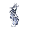 1bxxS S: Starting model for refinement |
|---|---|
| Similar structure data |


-Links
 PDBj
PDBj


- Assembly
| Deposited unit | 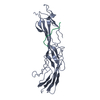
| ||||||||
|---|---|---|---|---|---|---|---|---|---|
| 1 |
| ||||||||
| Unit cell |
|
-Components
| #1: Protein | Mass: 33014.516 Da / Num. of mol.: 1 / Fragment: INTERNALIZATION SIGNAL BINDING DOMAIN Source method: isolated from a genetically manipulated source Details: THE FIRST 16 RESIDUES OF THE POLYMER MAKE UP A 6HIS-CMYC-TAG (HHHHHHEQKLISEEDL) Source: (gene. exp.) HOMO SAPIENS (human) / Production host:  |
|---|---|
| #2: Protein/peptide | Mass: 1194.419 Da / Num. of mol.: 1 / Source method: obtained synthetically Details: 11-MER INTERNALISATION SIGNAL MOTIF FROM CTLA-4 (HOMO SAPIENS), TTGVYVKMPPT Source: (synth.) HOMO SAPIENS (human) / References: UniProt: P16410 |
-Experimental details
-Experiment
| Experiment | Method: X-RAY DIFFRACTION / Number of used crystals: 1 |
|---|
- Sample preparation
| Crystal | Density Matthews: 5.03 Å3/Da / Density % sol: 75 % / Description: ISOMORPHOUS TO 1BXX | |||||||||||||||||||||||||||||||||||||||||||||||||||||||||||||||
|---|---|---|---|---|---|---|---|---|---|---|---|---|---|---|---|---|---|---|---|---|---|---|---|---|---|---|---|---|---|---|---|---|---|---|---|---|---|---|---|---|---|---|---|---|---|---|---|---|---|---|---|---|---|---|---|---|---|---|---|---|---|---|---|---|
| Crystal grow | Method: vapor diffusion, hanging drop / pH: 7 Details: HANGING DROPS CONTAINING A 1:1 MIXTURE OF COMPLEX SOLUTION 6MG/ML PROTEIN AND 1MM 11MER PEPTIDE SOLUTION IN 20MM HEPES PH7.5, 1MM DTT AND RESERVOIR BUFFER 1.4-2.4M SODIUM CHLORIDE, 0.1M MES ...Details: HANGING DROPS CONTAINING A 1:1 MIXTURE OF COMPLEX SOLUTION 6MG/ML PROTEIN AND 1MM 11MER PEPTIDE SOLUTION IN 20MM HEPES PH7.5, 1MM DTT AND RESERVOIR BUFFER 1.4-2.4M SODIUM CHLORIDE, 0.1M MES PH6.2-7.0, 0.4M NA/K PHOSPHATE, 15% GLYCEROL, pH 7.00 | |||||||||||||||||||||||||||||||||||||||||||||||||||||||||||||||
| Crystal grow | *PLUS Temperature: 15 ℃ / pH: 7.5 / Method: vapor diffusion | |||||||||||||||||||||||||||||||||||||||||||||||||||||||||||||||
| Components of the solutions | *PLUS
|
-Data collection
| Diffraction | Mean temperature: 100 K |
|---|---|
| Diffraction source | Source: SYNCHROTRON / Site: SRS  / Beamline: PX9.6 / Wavelength: 0.87 / Beamline: PX9.6 / Wavelength: 0.87 |
| Detector | Type: ADSC CCD / Detector: CCD / Date: Apr 26, 1999 / Details: MIRROR |
| Radiation | Monochromator: YES / Protocol: SINGLE WAVELENGTH / Monochromatic (M) / Laue (L): M / Scattering type: x-ray |
| Radiation wavelength | Wavelength: 0.87 Å / Relative weight: 1 |
| Reflection | Resolution: 3.6→48.2 Å / Num. obs: 6681 / % possible obs: 83.2 % / Redundancy: 2.2 % / Biso Wilson estimate: 0 Å2 / Rmerge(I) obs: 0.133 / Net I/σ(I): 4 |
| Reflection shell | Resolution: 3.6→3.8 Å / Redundancy: 2.1 % / Rmerge(I) obs: 0.354 / Mean I/σ(I) obs: 1.7 / % possible all: 85.9 |
| Reflection | *PLUS Num. measured all: 14633 |
| Reflection shell | *PLUS % possible obs: 85.9 % / Num. unique obs: 992 / Num. measured obs: 2081 |
- Processing
| Software |
| ||||||||||||||||||||||||||||||||||||||||||||||||||||||||||||
|---|---|---|---|---|---|---|---|---|---|---|---|---|---|---|---|---|---|---|---|---|---|---|---|---|---|---|---|---|---|---|---|---|---|---|---|---|---|---|---|---|---|---|---|---|---|---|---|---|---|---|---|---|---|---|---|---|---|---|---|---|---|
| Refinement | Method to determine structure: FOURIER SYNTHESIS Starting model: PDB ENTRY 1BXX Resolution: 3.6→7.99 Å / Rfactor Rfree error: 0.013 / Isotropic thermal model: GROUP / Cross valid method: THROUGHOUT / σ(F): 0
| ||||||||||||||||||||||||||||||||||||||||||||||||||||||||||||
| Solvent computation | Solvent model: FLAT MODEL / Bsol: 10 Å2 / ksol: 0.14399 e/Å3 | ||||||||||||||||||||||||||||||||||||||||||||||||||||||||||||
| Displacement parameters | Biso mean: 39.6 Å2
| ||||||||||||||||||||||||||||||||||||||||||||||||||||||||||||
| Refine analyze |
| ||||||||||||||||||||||||||||||||||||||||||||||||||||||||||||
| Refinement step | Cycle: LAST / Resolution: 3.6→7.99 Å
| ||||||||||||||||||||||||||||||||||||||||||||||||||||||||||||
| Refine LS restraints |
| ||||||||||||||||||||||||||||||||||||||||||||||||||||||||||||
| LS refinement shell | Resolution: 3.6→3.8 Å / Rfactor Rfree error: 0.067 / Total num. of bins used: 6
| ||||||||||||||||||||||||||||||||||||||||||||||||||||||||||||
| Xplor file | Serial no: 1 / Param file: PROTEIN_REP.PARAM / Topol file: PROTEIN.TOP | ||||||||||||||||||||||||||||||||||||||||||||||||||||||||||||
| Refinement | *PLUS Lowest resolution: 8 Å | ||||||||||||||||||||||||||||||||||||||||||||||||||||||||||||
| Solvent computation | *PLUS | ||||||||||||||||||||||||||||||||||||||||||||||||||||||||||||
| Displacement parameters | *PLUS | ||||||||||||||||||||||||||||||||||||||||||||||||||||||||||||
| Refine LS restraints | *PLUS
|
