[English] 日本語
Yorodumi
- PDB-1h2n: Factor Inhibiting HIF-1 alpha -

+
Open data


ID or keywords:

Loading...

-
Basic information

Entry
Database: PDB / ID: 1h2n
TitleFactor Inhibiting HIF-1 alpha
ComponentsHYPOXIA-INDUCIBLE FACTOR 1-ALPHA INHIBITOR
KeywordsOXIDOREDUCTASE / TRANSCRIPTION ACTIVATOR / OXYGENASE / HYPOXIA / HYDROXYLASE
Function / homology
Function and homology information


hypoxia-inducible factor-asparagine dioxygenase / : / [protein]-asparagine 3-dioxygenase activity / peptidyl-histidine dioxygenase activity / peptidyl-aspartic acid 3-dioxygenase activity / Cellular response to hypoxia / carboxylic acid binding / positive regulation of vasculogenesis / ankyrin repeat binding / oxygen sensor activity ...hypoxia-inducible factor-asparagine dioxygenase / : / [protein]-asparagine 3-dioxygenase activity / peptidyl-histidine dioxygenase activity / peptidyl-aspartic acid 3-dioxygenase activity / Cellular response to hypoxia / carboxylic acid binding / positive regulation of vasculogenesis / ankyrin repeat binding / oxygen sensor activity / Notch binding / negative regulation of Notch signaling pathway / NF-kappaB binding / positive regulation of myoblast differentiation / ferrous iron binding / transcription corepressor activity / perinuclear region of cytoplasm / protein homodimerization activity / zinc ion binding / nucleoplasm / cytoplasm / cytosol
Similarity search - Function
Clavaminate synthase-like / Hypoxia-inducible factor 1-alpha inhibitor, domain II / Cupin-like domain 8 / Cupin-like domain / A domain family that is part of the cupin metalloenzyme superfamily. / Jelly Rolls / JmjC domain / JmjC domain profile. / RmlC-like jelly roll fold / Helix Hairpins ...Clavaminate synthase-like / Hypoxia-inducible factor 1-alpha inhibitor, domain II / Cupin-like domain 8 / Cupin-like domain / A domain family that is part of the cupin metalloenzyme superfamily. / Jelly Rolls / JmjC domain / JmjC domain profile. / RmlC-like jelly roll fold / Helix Hairpins / Jelly Rolls / Sandwich / Orthogonal Bundle / Mainly Beta / Mainly Alpha
Similarity search - Domain/homology
2-OXOGLUTARIC ACID / : / Hypoxia-inducible factor 1-alpha inhibitor
Similarity search - Component
Biological speciesHOMO SAPIENS (human)
MethodX-RAY DIFFRACTION / SYNCHROTRON / MOLECULAR REPLACEMENT / Resolution: 2.84 Å
AuthorsElkins, J.M. / Hewitson, K.S. / McNeill, L.A. / Schlemminger, I. / Seibel, J.F. / Schofield, C.J.
CitationJournal: J.Biol.Chem. / Year: 2003
Title: Structure of Factor-Inhibiting Hypoxia-Inducible Factor (Hif) Reveals Mechanism of Oxidative Modification of Hif-1Alpha
Authors: Elkins, J.M. / Hewitson, K.S. / McNeill, L.A. / Seibel, J.F. / Schlemminger, I. / Pugh, C. / Ratcliffe, P. / Schofield, C.J.
History
DepositionAug 12, 2002Deposition site: PDBE / Processing site: PDBE
Revision 1.0Nov 28, 2002Provider: repository / Type: Initial release
Revision 1.1Jul 13, 2011Group: Advisory / Version format compliance
Revision 1.2Feb 21, 2018Group: Database references / Category: citation / citation_author / Item: _citation.page_last / _citation_author.name
Revision 1.3May 8, 2024Group: Data collection / Database references ...Data collection / Database references / Derived calculations / Other
Category: chem_comp_atom / chem_comp_bond ...chem_comp_atom / chem_comp_bond / database_2 / pdbx_database_status / struct_conn / struct_site
Item: _database_2.pdbx_DOI / _database_2.pdbx_database_accession ..._database_2.pdbx_DOI / _database_2.pdbx_database_accession / _pdbx_database_status.status_code_sf / _struct_conn.ptnr1_auth_comp_id / _struct_conn.ptnr1_auth_seq_id / _struct_conn.ptnr1_label_asym_id / _struct_conn.ptnr1_label_atom_id / _struct_conn.ptnr1_label_comp_id / _struct_conn.ptnr1_label_seq_id / _struct_conn.ptnr2_auth_comp_id / _struct_conn.ptnr2_auth_seq_id / _struct_conn.ptnr2_label_asym_id / _struct_conn.ptnr2_label_atom_id / _struct_conn.ptnr2_label_comp_id / _struct_conn.ptnr2_label_seq_id / _struct_site.pdbx_auth_asym_id / _struct_site.pdbx_auth_comp_id / _struct_site.pdbx_auth_seq_id
Remark 700 
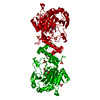
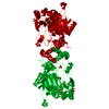
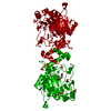
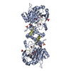
SHEET THE SHEET STRUCTURE OF THIS MOLECULE IS BIFURCATED. IN ORDER TO REPRESENT THIS FEATURE IN ... SHEET THE SHEET STRUCTURE OF THIS MOLECULE IS BIFURCATED. IN ORDER TO REPRESENT THIS FEATURE IN THE SHEET RECORDS BELOW, TWO SHEETS ARE DEFINED.

-
Structure visualization

Structure viewerMolecule:
MolmilJmol/JSmol

Downloads & links

-
Assembly

Deposited unit
A: HYPOXIA-INDUCIBLE FACTOR 1-ALPHA INHIBITOR
hetero molecules


Theoretical massNumber of molelcules
Total (without water)40,8186
Polymers40,3281
Non-polymers4905
Water543
1
A: HYPOXIA-INDUCIBLE FACTOR 1-ALPHA INHIBITOR
hetero molecules

A: HYPOXIA-INDUCIBLE FACTOR 1-ALPHA INHIBITOR
hetero molecules


Theoretical massNumber of molelcules
Total (without water)81,63712
Polymers80,6572
Non-polymers98010
Water362
TypeNameSymmetry operationNumber
identity operation1_555x,y,z1
crystal symmetry operation8_665-y+1,-x+1,-z+1/21
Buried area5320 Å2
ΔGint-131.55 kcal/mol
Surface area29910 Å2
MethodPISA
Unit cell
Length a, b, c (Å)86.342, 86.342, 146.732
Angle α, β, γ (deg.)90.00, 90.00, 90.00
Int Tables number92
Space group name H-MP41212
DetailsTHE PROTEIN IS A DIMER FORMED BY CHAIN A.

-
Components

#1: Protein HYPOXIA-INDUCIBLE FACTOR 1-ALPHA INHIBITOR / HYPOXIA-INDUCIBLE FACTOR ASPARAGINE HYDROXYLASE / FACTOR INHIBITING HIF-1 / FIH1


Mass: 40328.281 Da / Num. of mol.: 1
Source method: isolated from a genetically manipulated source
Source: (gene. exp.) HOMO SAPIENS (human) / Plasmid: PET28A(+) / Production host: ESCHERICHIA COLI (E. coli) / Strain (production host): BL21(DE3)
References: UniProt: Q9NWT6, peptide-aspartate beta-dioxygenase
#2: Chemical ChemComp-FE2 / FE (II) ION


Mass: 55.845 Da / Num. of mol.: 1 / Source method: obtained synthetically / Formula: Fe
#3: Chemical ChemComp-AKG / 2-OXOGLUTARIC ACID


Mass: 146.098 Da / Num. of mol.: 1 / Source method: obtained synthetically / Formula: C5H6O5
#4: Chemical ChemComp-SO4 / SULFATE ION


Mass: 96.063 Da / Num. of mol.: 3 / Source method: obtained synthetically / Formula: SO4
#5: Water ChemComp-HOH / water


Mass: 18.015 Da / Num. of mol.: 3 / Source method: isolated from a natural source / Formula: H2O
Compound detailsTHE PROTEIN (CHAIN A) WAS CRYSTALLIZED IN THE PRESENCE OF A PEPTIDE FRAGEMENT FROM ENDOTHELIAL PAS ...THE PROTEIN (CHAIN A) WAS CRYSTALLIZED IN THE PRESENCE OF A PEPTIDE FRAGEMENT FROM ENDOTHELIAL PAS DOMAIN PROTEIN 1 SWISS-PROT ID Q99814 (RESIDUES 846-858) BUT NONE OF THE RESIDUES CORRESPONDING TO THE PEPTIDE WERE VISIBLE IN THE ELECTRON DENSITY MAPS. IT IS POSSIBLE THAT THE PEPTIDE DID NOT BIND TO THE PROTEIN AND HENCE HAS NOT BEEN INCLUDED IN THE COMPND, SOURCE AND SEQRES RECORDS. THE SEQUENCE OF THE FRAGMENT IS GIVEN BELOW. VAL ASN VAL PRO VAL LEU GLY SER SER THR LEU LEU GLN

-
Experimental details

-
Experiment

ExperimentMethod: X-RAY DIFFRACTION / Number of used crystals: 1

-
Sample preparation

CrystalDensity Matthews: 3.4 Å3/Da / Density % sol: 63 % / Description: SEE REMARK 400
Crystal growpH: 7.5
Details: 1.2M AMMONIUM SULPHATE, 4% PEG400, 0.1M HEPES PH7.5 ARGON ATMOSPHERE, 11MG/ML PROTEIN WITH 1MM FE(II), 2.5MM AKG AND 2.5MM PEPTIDE (SEE REMARK 400), pH 7.50
Crystal grow
*PLUS
Temperature: 17 ℃ / Method: vapor diffusion, hanging drop
Components of the solutions
*PLUS
IDConc.Common nameCrystal-IDSol-IDDetails
11.2 Mammonium sulfate1reservoir
24 %PEG4001reservoir
30.1 MHEPES1reservoirpH7.5
411 mg/mlFIH1drop
51 mMFe2+1drop
62 mM2OG/NOG1drop
71 mMCAD fragment1drop

-
Data collection

DiffractionMean temperature: 100 K
Diffraction sourceSource: SYNCHROTRON / Site: SRS / Beamline: PX9.5 / Wavelength: 0.92
DetectorType: MARRESEARCH / Detector: CCD / Date: May 15, 2002
RadiationProtocol: SINGLE WAVELENGTH / Monochromatic (M) / Laue (L): M / Scattering type: x-ray
Radiation wavelengthWavelength: 0.92 Å / Relative weight: 1
ReflectionResolution: 2.84→34.1 Å / Num. obs: 13703 / % possible obs: 99.2 % / Redundancy: 6.7 % / Rmerge(I) obs: 0.067 / Net I/σ(I): 9.4
Reflection shellResolution: 2.84→2.99 Å / Redundancy: 5.2 % / Rmerge(I) obs: 0.309 / Mean I/σ(I) obs: 2.4 / % possible all: 94.9

-
Processing

Software
NameVersionClassification
REFMAC5refinement
MOSFLMdata reduction
SCALAdata scaling
RefinementMethod to determine structure: MOLECULAR REPLACEMENT / Resolution: 2.84→30 Å / Cor.coef. Fo:Fc: 0.913 / Cor.coef. Fo:Fc free: 0.901 / SU B: 19.679 / SU ML: 0.398 / TLS residual ADP flag: LIKELY RESIDUAL / Cross valid method: THROUGHOUT / ESU R: 0.852 / ESU R Free: 0.349 / Stereochemistry target values: MAXIMUM LIKELIHOOD / Details: SEE REMARK 400
RfactorNum. reflection% reflectionSelection details
Rfree0.257 1046 7.7 %RANDOM
Rwork0.231 ---
obs0.233 12577 99.5 %-
Solvent computationIon probe radii: 0.8 Å / Shrinkage radii: 0.8 Å / VDW probe radii: 1.4 Å / Solvent model: BABINET MODEL WITH MASK
Displacement parametersBiso mean: 35.34 Å2
Baniso -1Baniso -2Baniso -3
1--1.02 Å20 Å20 Å2
2---1.02 Å20 Å2
3---2.03 Å2
Refinement stepCycle: LAST / Resolution: 2.84→30 Å
ProteinNucleic acidLigandSolventTotal
Num. atoms2689 0 26 3 2718
Refine LS restraints
Refine-IDTypeDev idealDev ideal targetNumber
X-RAY DIFFRACTIONr_bond_refined_d0.0150.0212791
X-RAY DIFFRACTIONr_bond_other_d0.0010.022388
X-RAY DIFFRACTIONr_angle_refined_deg1.6281.9453799
X-RAY DIFFRACTIONr_angle_other_deg0.82335576
X-RAY DIFFRACTIONr_dihedral_angle_1_deg4.2683330
X-RAY DIFFRACTIONr_dihedral_angle_2_deg
X-RAY DIFFRACTIONr_dihedral_angle_3_deg18.08215479
X-RAY DIFFRACTIONr_dihedral_angle_4_deg
X-RAY DIFFRACTIONr_chiral_restr0.0950.2384
X-RAY DIFFRACTIONr_gen_planes_refined0.0050.023137
X-RAY DIFFRACTIONr_gen_planes_other0.0020.02575
X-RAY DIFFRACTIONr_nbd_refined0.2550.3717
X-RAY DIFFRACTIONr_nbd_other0.2240.32425
X-RAY DIFFRACTIONr_nbtor_refined
X-RAY DIFFRACTIONr_nbtor_other
X-RAY DIFFRACTIONr_xyhbond_nbd_refined0.1390.5165
X-RAY DIFFRACTIONr_xyhbond_nbd_other0.1020.51
X-RAY DIFFRACTIONr_metal_ion_refined0.1120.53
X-RAY DIFFRACTIONr_metal_ion_other
X-RAY DIFFRACTIONr_symmetry_vdw_refined0.2560.314
X-RAY DIFFRACTIONr_symmetry_vdw_other0.2730.362
X-RAY DIFFRACTIONr_symmetry_hbond_refined0.2140.54
X-RAY DIFFRACTIONr_symmetry_hbond_other0.0610.51
X-RAY DIFFRACTIONr_symmetry_metal_ion_refined
X-RAY DIFFRACTIONr_symmetry_metal_ion_other
X-RAY DIFFRACTIONr_mcbond_it0.3121.51659
X-RAY DIFFRACTIONr_mcbond_other
X-RAY DIFFRACTIONr_mcangle_it0.59822675
X-RAY DIFFRACTIONr_mcangle_other
X-RAY DIFFRACTIONr_scbond_it1.05831132
X-RAY DIFFRACTIONr_scbond_other
X-RAY DIFFRACTIONr_scangle_it1.7954.51124
X-RAY DIFFRACTIONr_scangle_other
X-RAY DIFFRACTIONr_long_range_B_refined
X-RAY DIFFRACTIONr_long_range_B_other
X-RAY DIFFRACTIONr_rigid_bond_restr
X-RAY DIFFRACTIONr_sphericity_free
X-RAY DIFFRACTIONr_sphericity_bonded
LS refinement shellResolution: 2.84→2.91 Å / Total num. of bins used: 20 /
RfactorNum. reflection
Rfree0.315 81
Rwork0.286 828
Refinement TLS params.Method: refined / Origin x: 21.449 Å / Origin y: 27.42 Å / Origin z: 27.787 Å
111213212223313233
T0.223 Å20.0111 Å2-0.0923 Å2-0.0562 Å20.0525 Å2--0.0967 Å2
L1.6842 °21.5597 °21.1572 °2-4.4489 °22.3523 °2--2.0658 °2
S0.1098 Å °-0.2106 Å °-0.0766 Å °0.3449 Å °-0.0455 Å °0.2455 Å °0.3515 Å °-0.1199 Å °-0.0643 Å °
Refinement
*PLUS
Rfactor Rfree: 0.257 / Rfactor Rwork: 0.231
Solvent computation
*PLUS
Displacement parameters
*PLUS

+
About Yorodumi

-
News

-
Feb 9, 2022. New format data for meta-information of EMDB entries

New format data for meta-information of EMDB entries

  • Version 3 of the EMDB header file is now the official format.
  • The previous official version 1.9 will be removed from the archive.

Related info.:EMDB header

External links:wwPDB to switch to version 3 of the EMDB data model

-
Aug 12, 2020. Covid-19 info

Covid-19 info

URL: https://pdbjlvh1.pdbj.org/emnavi/covid19.php

New page: Covid-19 featured information page in EM Navigator.

Related info.:Covid-19 info / Mar 5, 2020. Novel coronavirus structure data

+
Mar 5, 2020. Novel coronavirus structure data

Novel coronavirus structure data

Related info.:Yorodumi Speices / Aug 12, 2020. Covid-19 info

External links:COVID-19 featured content - PDBj / Molecule of the Month (242):Coronavirus Proteases

+
Jan 31, 2019. EMDB accession codes are about to change! (news from PDBe EMDB page)

EMDB accession codes are about to change! (news from PDBe EMDB page)

  • The allocation of 4 digits for EMDB accession codes will soon come to an end. Whilst these codes will remain in use, new EMDB accession codes will include an additional digit and will expand incrementally as the available range of codes is exhausted. The current 4-digit format prefixed with “EMD-” (i.e. EMD-XXXX) will advance to a 5-digit format (i.e. EMD-XXXXX), and so on. It is currently estimated that the 4-digit codes will be depleted around Spring 2019, at which point the 5-digit format will come into force.
  • The EM Navigator/Yorodumi systems omit the EMD- prefix.

Related info.:Q: What is EMD? / ID/Accession-code notation in Yorodumi/EM Navigator

External links:EMDB Accession Codes are Changing Soon! / Contact to PDBj

+
Jul 12, 2017. Major update of PDB

Major update of PDB

  • wwPDB released updated PDB data conforming to the new PDBx/mmCIF dictionary.
  • This is a major update changing the version number from 4 to 5, and with Remediation, in which all the entries are updated.
  • In this update, many items about electron microscopy experimental information are reorganized (e.g. em_software).
  • Now, EM Navigator and Yorodumi are based on the updated data.

External links:wwPDB Remediation / Enriched Model Files Conforming to OneDep Data Standards Now Available in the PDB FTP Archive

-
Yorodumi

Thousand views of thousand structures

  • Yorodumi is a browser for structure data from EMDB, PDB, SASBDB, etc.
  • This page is also the successor to EM Navigator detail page, and also detail information page/front-end page for Omokage search.
  • The word "yorodu" (or yorozu) is an old Japanese word meaning "ten thousand". "mi" (miru) is to see.

Related info.:EMDB / PDB / SASBDB / Comparison of 3 databanks / Yorodumi Search / Aug 31, 2016. New EM Navigator & Yorodumi / Yorodumi Papers / Jmol/JSmol / Function and homology information / Changes in new EM Navigator and Yorodumi

Read more