 Movie
Movie Controller

[English] 日本語
 Yorodumi
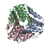
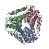
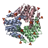
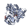
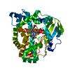
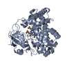
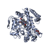
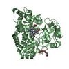
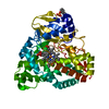
- PDB-1gu9: Crystal Structure of Mycobacterium tuberculosis Alkylperoxidase AhpD -
+ Open data

- Basic information
| Entry | Database: PDB / ID: 1gu9 | ||||||
|---|---|---|---|---|---|---|---|
| Title | Crystal Structure of Mycobacterium tuberculosis Alkylperoxidase AhpD | ||||||
 Components Components | ALKYLHYDROPEROXIDASE D | ||||||
 Keywords Keywords | OXIDOREDUCTASE / ALKYLHYDROPEROXIDASE / TUBERCULOSIS | ||||||
| Function / homology | AhpD-like / AhpD-like / Up-down Bundle / Mainly Alpha Function and homology information Function and homology information | ||||||
| Biological species |   MYCOBACTERIUM TUBERCULOSIS (bacteria) MYCOBACTERIUM TUBERCULOSIS (bacteria) | ||||||
| Method |  X-RAY DIFFRACTION / SYNCHROTRON / MAD / Resolution: 1.9 Å X-RAY DIFFRACTION / SYNCHROTRON / MAD / Resolution: 1.9 Å | ||||||
 Authors Authors | Nunn, C.M. / Djordjevic, S. / Hillas, P.J. / Nishida, C. / Ortiz de Montellano, P.R. | ||||||
 Citation Citation | Journal: J.Biol.Chem. / Year: 2002 Title: The Crystal Structure of Mycobacterium Tuberculosis Alkylhydroperoxidase Ahpd, a Potential Target for Antitubercular Drug Design Authors: Nunn, C.M. / Djordjevic, S. / Hillas, P.J. / Nishida, C. / Ortiz de Montellano, P.R. | ||||||
| History |
|
- Structure visualization
| Structure viewer | Molecule: MolmilJmol/JSmol |
|---|
- Downloads & links
-Download
| PDBx/mmCIF format | 1gu9.cif.gz | 393.7 KB | Display | PDBx/mmCIF format |
|---|---|---|---|---|
| PDB format | pdb1gu9.ent.gz | 327.6 KB | Display | PDB format |
| PDBx/mmJSON format | 1gu9.json.gz | Tree view | PDBx/mmJSON format | |
| Others |  Other downloads Other downloads |
-Validation report
| Arichive directory | https://data.pdbj.org/pub/pdb/validation_reports/gu/1gu9ftp://data.pdbj.org/pub/pdb/validation_reports/gu/1gu9 | HTTPS FTP |
|---|
-Related structure data
| Similar structure data |
|---|

-Links
 PDBj
PDBj- Assembly
| Deposited unit | 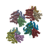
| ||||||||
|---|---|---|---|---|---|---|---|---|---|
| 1 | 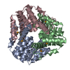
| ||||||||
| 2 | 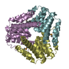
| ||||||||
| 3 | 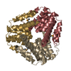
| ||||||||
| 4 | 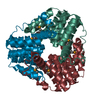
| ||||||||
| Unit cell |
|
-Components
| #1: Protein | Mass: 18942.205 Da / Num. of mol.: 12 Source method: isolated from a genetically manipulated source Source: (gene. exp.) MYCOBACTERIUM TUBERCULOSIS (bacteria) / Strain: H37RV / Production host: #2: Water | ChemComp-HOH / |  Mass: 18.015 Da / Num. of mol.: 847 / Source method: isolated from a natural source / Formula: H2O Mass: 18.015 Da / Num. of mol.: 847 / Source method: isolated from a natural source / Formula: H2OHas protein modification | Y | Sequence details | RESIDUE 104 IN ALL CHAINS WAS FOUND TO BE A METHIONINE | |
|---|
-Experimental details
-Experiment
| Experiment | Method: X-RAY DIFFRACTION / Number of used crystals: 1 |
|---|
- Sample preparation
| Crystal | Density Matthews: 1.97 Å3/Da / Density % sol: 37 % | ||||||||||||||||||||||||||||||||||||||||||||||||||||||||||||||||||||||
|---|---|---|---|---|---|---|---|---|---|---|---|---|---|---|---|---|---|---|---|---|---|---|---|---|---|---|---|---|---|---|---|---|---|---|---|---|---|---|---|---|---|---|---|---|---|---|---|---|---|---|---|---|---|---|---|---|---|---|---|---|---|---|---|---|---|---|---|---|---|---|---|
| Crystal grow | pH: 6 Details: 100MM SODIUM CITRATE BUFFER, PH 5.6, CONTAINING 200MM AMMONIUM ACETATE AND 26% PEG 4000. MIXED IN EQUAL VOLUME WITH AHPD (4.5 MG/ML) IN 25 MM MOPS BUFFER, PH 7.2, CONTAINING 50 MM KCL, 10% GLYCEROL, 0.1MM EDTA | ||||||||||||||||||||||||||||||||||||||||||||||||||||||||||||||||||||||
| Crystal grow | *PLUS Temperature: 4 ℃ / pH: 7.2 / Method: vapor diffusion, hanging drop | ||||||||||||||||||||||||||||||||||||||||||||||||||||||||||||||||||||||
| Components of the solutions | *PLUS
|
-Data collection
| Diffraction | Mean temperature: 110 K | |||||||||||||||
|---|---|---|---|---|---|---|---|---|---|---|---|---|---|---|---|---|
| Diffraction source | Source: SYNCHROTRON / Site: ESRF  / Beamline: BM14 / Wavelength: 0.88550,0.918400,0.978900 , 0.97877 / Beamline: BM14 / Wavelength: 0.88550,0.918400,0.978900 , 0.97877 | |||||||||||||||
| Detector | Type: MARRESEARCH / Detector: CCD / Date: Jun 15, 2001 / Details: MIRRORS | |||||||||||||||
| Radiation | Monochromator: SI 111 / Protocol: MAD / Monochromatic (M) / Laue (L): M / Scattering type: x-ray | |||||||||||||||
| Radiation wavelength |
| |||||||||||||||
| Reflection | Resolution: 1.9→30 Å / Num. obs: 136927 / % possible obs: 99.5 % / Observed criterion σ(I): 2 / Redundancy: 3.7 % / Biso Wilson estimate: 19.1 Å2 / Rmerge(I) obs: 0.072 / Net I/σ(I): 7.2 | |||||||||||||||
| Reflection shell | Resolution: 1.9→1.97 Å / Redundancy: 3.4 % / Rmerge(I) obs: 0.42 / Mean I/σ(I) obs: 2.75 / % possible all: 99.3 | |||||||||||||||
| Reflection | *PLUS Lowest resolution: 500 Å / Num. measured all: 1263412 | |||||||||||||||
| Reflection shell | *PLUS % possible obs: 99.3 % |
- Processing
| Software |
| ||||||||||||||||||||||||||||||||||||||||||||||||||||||||||||||||||||||||||||||||
|---|---|---|---|---|---|---|---|---|---|---|---|---|---|---|---|---|---|---|---|---|---|---|---|---|---|---|---|---|---|---|---|---|---|---|---|---|---|---|---|---|---|---|---|---|---|---|---|---|---|---|---|---|---|---|---|---|---|---|---|---|---|---|---|---|---|---|---|---|---|---|---|---|---|---|---|---|---|---|---|---|---|
| Refinement | Method to determine structure: MAD / Resolution: 1.9→29.6 Å / Rfactor Rfree error: 0.006 / Isotropic thermal model: RESTRAINED / Cross valid method: THROUGHOUT / σ(F): 2
| ||||||||||||||||||||||||||||||||||||||||||||||||||||||||||||||||||||||||||||||||
| Solvent computation | Solvent model: FLAT MODEL / Bsol: 57.9056 Å2 / ksol: 0.349187 e/Å3 | ||||||||||||||||||||||||||||||||||||||||||||||||||||||||||||||||||||||||||||||||
| Displacement parameters | Biso mean: 33.8 Å2
| ||||||||||||||||||||||||||||||||||||||||||||||||||||||||||||||||||||||||||||||||
| Refine analyze |
| ||||||||||||||||||||||||||||||||||||||||||||||||||||||||||||||||||||||||||||||||
| Refinement step | Cycle: LAST / Resolution: 1.9→29.6 Å
| ||||||||||||||||||||||||||||||||||||||||||||||||||||||||||||||||||||||||||||||||
| Refine LS restraints |
| ||||||||||||||||||||||||||||||||||||||||||||||||||||||||||||||||||||||||||||||||
| LS refinement shell | Resolution: 1.9→1.97 Å / Rfactor Rfree error: 0.008 / Total num. of bins used: 10
| ||||||||||||||||||||||||||||||||||||||||||||||||||||||||||||||||||||||||||||||||
| Xplor file |
| ||||||||||||||||||||||||||||||||||||||||||||||||||||||||||||||||||||||||||||||||
| Software | *PLUS Name: CNS / Version: 1 / Classification: refinement | ||||||||||||||||||||||||||||||||||||||||||||||||||||||||||||||||||||||||||||||||
| Refinement | *PLUS Lowest resolution: 500 Å / Rfactor Rfree: 0.31 | ||||||||||||||||||||||||||||||||||||||||||||||||||||||||||||||||||||||||||||||||
| Solvent computation | *PLUS | ||||||||||||||||||||||||||||||||||||||||||||||||||||||||||||||||||||||||||||||||
| Displacement parameters | *PLUS Biso mean: 33.4 Å2 | ||||||||||||||||||||||||||||||||||||||||||||||||||||||||||||||||||||||||||||||||
| Refine LS restraints | *PLUS
|
