[English] 日本語
Yorodumi
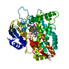
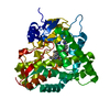
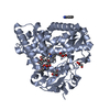
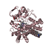
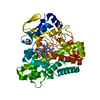
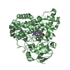
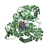
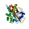
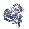
- PDB-4z5p: Crystal structure of the LnmA cytochrome P450 hydroxylase from th... -

+
Open data


ID or keywords:

Loading...

-
Basic information

Entry
Database: PDB / ID: 4z5p
TitleCrystal structure of the LnmA cytochrome P450 hydroxylase from the leinamycin biosynthetic pathway of Streptomyces atroolivaceus S-140 at 1.9 A resolution
ComponentsCytochrome P450 hydroxylase
KeywordsOXIDOREDUCTASE / hydroxylase / leinamycin / heme / monooxygenase / Structural Genomics / PSI-Biology / Midwest Center for Structural Genomics / MCSG / Enzyme Discovery for Natural Product Biosynthesis / NatPro
Function / homology
Function and homology information


oxidoreductase activity, acting on paired donors, with incorporation or reduction of molecular oxygen / monooxygenase activity / iron ion binding / heme binding
Similarity search - Function
Cytochrome P450, B-class / Cytochrome p450 / Cytochrome P450 / Cytochrome P450, conserved site / Cytochrome P450 cysteine heme-iron ligand signature. / Cytochrome P450 / Cytochrome P450 superfamily / Cytochrome P450 / Orthogonal Bundle / Mainly Alpha
Similarity search - Domain/homology
PROTOPORPHYRIN IX CONTAINING FE / TRIETHYLENE GLYCOL / Cytochrome P450 hydroxylase
Similarity search - Component
Biological speciesStreptomyces atroolivaceus (bacteria)
MethodX-RAY DIFFRACTION / SYNCHROTRON / MOLECULAR REPLACEMENT / molecular replacement / Resolution: 1.9 Å
AuthorsMa, M. / Lohman, J. / Rudolf, J. / Miller, M.D. / Cao, H. / Osipiuk, J. / Babnigg, G. / Phillips Jr., G.N. / Joachimiak, A. / Shen, B. ...Ma, M. / Lohman, J. / Rudolf, J. / Miller, M.D. / Cao, H. / Osipiuk, J. / Babnigg, G. / Phillips Jr., G.N. / Joachimiak, A. / Shen, B. / Midwest Center for Structural Genomics (MCSG) / Enzyme Discovery for Natural Product Biosynthesis (NatPro)
Funding support United States, 2items
OrganizationGrant numberCountry
National Institutes of Health/National Institute of General Medical Sciences (NIH/NIGMS)U01GM098248 United States
National Institutes of Health/National Cancer Institute (NIH/NCI)R01CA106150 United States
CitationJournal: To be Published
Title: Crystal structure of the LnmA cytochrome P450 hydroxylase from the leinamycin biosynthetic pathway of Streptomyces atroolivaceus S-140
Authors: Ma, M. / Lohman, J. / Rudolf, J. / Miller, M.D. / Cao, H. / Osipiuk, J. / Babnigg, G. / Phillips Jr., G.N. / Joachimiak, A. / Shen, B.
History
DepositionApr 2, 2015Deposition site: RCSB / Processing site: RCSB
Revision 1.0Jul 29, 2015Provider: repository / Type: Initial release
Revision 1.1Sep 20, 2017Group: Author supporting evidence / Derived calculations ...Author supporting evidence / Derived calculations / Refinement description / Source and taxonomy
Category: entity_src_gen / pdbx_audit_support ...entity_src_gen / pdbx_audit_support / pdbx_struct_oper_list / software
Item: _entity_src_gen.pdbx_alt_source_flag / _pdbx_audit_support.funding_organization / _pdbx_struct_oper_list.symmetry_operation
Revision 1.2Dec 4, 2019Group: Author supporting evidence / Category: pdbx_audit_support / Item: _pdbx_audit_support.funding_organization
Revision 1.3Sep 27, 2023Group: Data collection / Database references ...Data collection / Database references / Derived calculations / Refinement description
Category: chem_comp_atom / chem_comp_bond ...chem_comp_atom / chem_comp_bond / database_2 / pdbx_initial_refinement_model / struct_conn
Item: _database_2.pdbx_DOI / _database_2.pdbx_database_accession ..._database_2.pdbx_DOI / _database_2.pdbx_database_accession / _struct_conn.pdbx_dist_value / _struct_conn.ptnr1_auth_asym_id / _struct_conn.ptnr1_auth_comp_id / _struct_conn.ptnr1_auth_seq_id / _struct_conn.ptnr1_label_asym_id / _struct_conn.ptnr1_label_atom_id / _struct_conn.ptnr1_label_comp_id / _struct_conn.ptnr1_label_seq_id / _struct_conn.ptnr2_auth_asym_id / _struct_conn.ptnr2_auth_comp_id / _struct_conn.ptnr2_auth_seq_id / _struct_conn.ptnr2_label_asym_id / _struct_conn.ptnr2_label_atom_id / _struct_conn.ptnr2_label_comp_id

-
Structure visualization

Structure viewerMolecule:
MolmilJmol/JSmol

Downloads & links

-
Assembly

Deposited unit
A: Cytochrome P450 hydroxylase
B: Cytochrome P450 hydroxylase
hetero molecules


Theoretical massNumber of molelcules
Total (without water)90,1525
Polymers88,7682
Non-polymers1,3833
Water6,828379
1
A: Cytochrome P450 hydroxylase
hetero molecules


Theoretical massNumber of molelcules
Total (without water)45,0012
Polymers44,3841
Non-polymers6161
Water181
TypeNameSymmetry operationNumber
identity operation1_555x,y,z1
2
B: Cytochrome P450 hydroxylase
hetero molecules


Theoretical massNumber of molelcules
Total (without water)45,1513
Polymers44,3841
Non-polymers7672
Water181
TypeNameSymmetry operationNumber
identity operation1_555x,y,z1
Unit cell
Length a, b, c (Å)55.759, 75.454, 93.858
Angle α, β, γ (deg.)90.000, 98.910, 90.000
Int Tables number4
Space group name H-MP1211

-
Components

#1: Protein Cytochrome P450 hydroxylase


Mass: 44384.188 Da / Num. of mol.: 2
Source method: isolated from a genetically manipulated source
Source: (gene. exp.) Streptomyces atroolivaceus (bacteria) / Strain: S-140 / Gene: LnmA / Plasmid: pRSF-TEV/LIC / Production host: Escherichia Coli (E. coli) / Strain (production host): BL21(DE3) / References: UniProt: Q8GGQ1
#2: Chemical ChemComp-HEM / PROTOPORPHYRIN IX CONTAINING FE / HEME


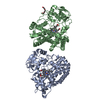
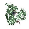
Mass: 616.487 Da / Num. of mol.: 2 / Source method: obtained synthetically / Formula: C34H32FeN4O4
#3: Chemical ChemComp-PGE / TRIETHYLENE GLYCOL


Mass: 150.173 Da / Num. of mol.: 1 / Source method: obtained synthetically / Formula: C6H14O4
#4: Water ChemComp-HOH / water


Mass: 18.015 Da / Num. of mol.: 379 / Source method: isolated from a natural source / Formula: H2O
Sequence detailsTHE CONSTRUCT WAS EXPRESSED WITH AN N-TERMINAL PURIFICATION TAG MGSSHHHHHHSQDPGDENLYFQS. THE TAG ...THE CONSTRUCT WAS EXPRESSED WITH AN N-TERMINAL PURIFICATION TAG MGSSHHHHHHSQDPGDENLYFQS. THE TAG WAS REMOVED WITH TEV PROTEASE LEAVING ONLY A SERINE (0) FOLLOWED BY THE TARGET SEQUENCE.

-
Experimental details

-
Experiment

ExperimentMethod: X-RAY DIFFRACTION / Number of used crystals: 1

-
Sample preparation

CrystalDensity Matthews: 2.2 Å3/Da / Density % sol: 44.02 %
Crystal growTemperature: 298 K / Method: vapor diffusion, sitting drop / pH: 6.77
Details: 0.2 M ammonium acetate, 0.1 M Bis-Tris, pH 6.77, 25% PEG3350, VAPOR DIFFUSION, SITTING DROP
Temp details: room temperature
Experiment crystal grow comp
ConcComp nameComp-IDCrystal-IDSol-ID
17. mg/mlprotein11macromolecule
10. mMTEAOH21macromolecule
20. mMNaCl31macromolecule
10. mMKCl41macromolecule
25. %(w/v)PEG 335051precipitant
0.1 MBis-Tris61precipitant
0.2 Mammonium acetate71precipitant
25. %(w/v)PEG 335081reservoir
0.1 MBis-Tris91reservoir
0.2 Mammonium acetate101reservoir
Experiment crystal grow sol
Crystal-IDSol-IDpHVolume3)
1macromolecule7.51.0 µL
1precipitant6.771.0 µL
1reservoir6.770.5 mL

-
Data collection

DiffractionMean temperature: 100 K / Crystal support: 0.2-0.3 mm nylon loop
Diffraction sourceSource: SYNCHROTRON / Site: APS / Beamline: 19-ID / Wavelength: 0.97918 Å
DetectorType: ADSC QUANTUM 315r / Detector: CCD / Date: Apr 10, 2014 / Details: mirrors
RadiationMonochromator: double crystals / Protocol: SINGLE WAVELENGTH / Monochromatic (M) / Laue (L): M / Scattering type: x-ray
Radiation wavelengthWavelength: 0.97918 Å / Relative weight: 1
ReflectionResolution: 1.9→46.36 Å / Num. all: 57513 / Num. obs: 57513 / % possible obs: 94.9 % / Redundancy: 3.7 % / CC1/2: 0.997 / Rpim(I) all: 0.056 / Rrim(I) all: 0.108 / Rsym value: 0.092 / Net I/av σ(I): 7.03 / Net I/σ(I): 11.1 / Num. measured all: 210829
Reflection shell
Resolution (Å)Redundancy (%)Rmerge(I) allRmerge(I) obsMean I/σ(I) obsNum. measured allNum. unique allCC1/2Rpim(I) allRrim(I) allRsym valueDiffraction-IDNet I/σ(I) obs% possible all
1.9-1.953.20.9910.8331.61007631150.7330.530.9910.83311.670.1
1.95-23.20.8420.70121255239160.7420.4580.8420.701290.5
2-2.063.40.6810.5742.81407741020.820.3620.6810.5742.897.1
2.06-2.123.80.5690.4883.61518039840.8490.290.5690.4883.697.5
2.12-2.193.80.4650.44.41494739100.8930.2370.4650.44.497.5
2.19-2.273.80.4030.3465.11441137620.9090.2050.4030.3465.197.6
2.27-2.363.80.3290.28261391336460.9350.1680.3290.282697.7
2.36-2.453.80.2780.23871335835030.9490.1420.2780.238798.2
2.45-2.563.80.2320.1997.91282833880.9670.1180.2320.1997.998.3
2.56-2.693.80.1830.1579.41227632400.9780.0940.1830.1579.497.8
2.69-2.833.80.150.12810.81157430600.9840.0770.150.12810.898.2
2.83-33.70.120.10212.81087029050.990.0620.120.10212.897.8
3-3.213.70.0870.07416.41014127110.9950.0450.0870.07416.497.5
3.21-3.473.70.0650.05520.3942725380.9970.0330.0650.05520.396.8
3.47-3.83.70.0490.04225.8842622910.9970.0260.0490.04225.896.1
3.8-4.253.70.0420.03629.8768821000.9980.0220.0420.03629.896.2
4.25-4.913.50.040.03432.3651218360.9980.0210.040.03432.396.4
4.91-6.013.60.0420.03628.9564015780.9980.0220.0420.03628.996.3
6.01-8.53.70.0380.03231.2453312410.9980.020.0380.03231.296.9
8.5-46.363.50.0330.02837.924006870.9980.0170.0330.02837.996

-
Phasing

PhasingMethod: molecular replacement

-
Processing

Software
NameVersionClassificationNB
XDSdata reduction
PHASERphasing
REFMAC5.8.0107refinement
PDB_EXTRACT3.11data extraction
SBC-Collectdata collection
XDSdata scaling
RefinementMethod to determine structure: MOLECULAR REPLACEMENT
Starting model: 4Z5Q
Resolution: 1.9→46.36 Å / Cor.coef. Fo:Fc: 0.957 / Cor.coef. Fo:Fc free: 0.945 / Occupancy max: 1 / Occupancy min: 0.4 / SU B: 9.452 / SU ML: 0.132 / Cross valid method: THROUGHOUT / σ(F): 0 / ESU R: 0.181 / ESU R Free: 0.148 / Stereochemistry target values: MAXIMUM LIKELIHOOD
Details: 1. HYDROGENS HAVE BEEN ADDED IN THE RIDING POSITIONS 2. ATOM RECORD CONTAINS SUM OF TLS AND RESIDUAL B FACTORS. ANISOU RECORD CONTAINS SUM OF TLS AND RESIDUAL U FACTORS. 3. WATERS WERE ...Details: 1. HYDROGENS HAVE BEEN ADDED IN THE RIDING POSITIONS 2. ATOM RECORD CONTAINS SUM OF TLS AND RESIDUAL B FACTORS. ANISOU RECORD CONTAINS SUM OF TLS AND RESIDUAL U FACTORS. 3. WATERS WERE EXCLUDED FROM AUTOMATIC TLS ASSIGNMENT. 4. THE HEME IRON IS MODELED AS FE2+ AND IS LIKELY REDUCED BY X-RAY EXPOSURE. 5. A PEG FRAGMENT FROM THE CRYSTLLIZATOION CONDITIONS HAVE BEEN MODELED.
RfactorNum. reflection% reflectionSelection details
Rfree0.2167 2884 5 %RANDOM
Rwork0.1908 54626 --
obs0.1922 57510 94.73 %-
Solvent computationIon probe radii: 0.8 Å / Shrinkage radii: 0.8 Å / VDW probe radii: 1.2 Å / Solvent model: MASK
Displacement parametersBiso max: 114.12 Å2 / Biso mean: 34.8009 Å2 / Biso min: 12.61 Å2
Baniso -1Baniso -2Baniso -3
1-0.8 Å20 Å20.6 Å2
2---0.23 Å20 Å2
3----0.72 Å2
Refinement stepCycle: LAST / Resolution: 1.9→46.36 Å
ProteinNucleic acidLigandSolventTotal
Num. atoms6005 0 96 379 6480
Refine LS restraints
Refine-IDTypeDev idealDev ideal targetNumber
X-RAY DIFFRACTIONr_bond_refined_d0.0120.0196359
X-RAY DIFFRACTIONr_bond_other_d0.0040.026061
X-RAY DIFFRACTIONr_angle_refined_deg1.491.9898689
X-RAY DIFFRACTIONr_angle_other_deg1.083313860
X-RAY DIFFRACTIONr_dihedral_angle_1_deg5.5255804
X-RAY DIFFRACTIONr_dihedral_angle_2_deg26.17422.391297
X-RAY DIFFRACTIONr_dihedral_angle_3_deg13.099151001
X-RAY DIFFRACTIONr_dihedral_angle_4_deg15.4021575
X-RAY DIFFRACTIONr_chiral_restr0.0930.2958
X-RAY DIFFRACTIONr_gen_planes_refined0.0070.0217295
X-RAY DIFFRACTIONr_gen_planes_other0.0050.021504
X-RAY DIFFRACTIONr_mcbond_it2.1953.8053150
X-RAY DIFFRACTIONr_mcbond_other2.1943.8053149
X-RAY DIFFRACTIONr_mcangle_it3.2987.1013946
LS refinement shellResolution: 1.9→1.95 Å / Total num. of bins used: 20
RfactorNum. reflection% reflection
Rfree0.332 143 -
Rwork0.316 2968 -
all-3111 -
obs--69.1 %
Refinement TLS params.

Method: refined / Refine-ID: X-RAY DIFFRACTION

IDL112)L122)L132)L222)L232)L332)S11 (Å °)S12 (Å °)S13 (Å °)S21 (Å °)S22 (Å °)S23 (Å °)S31 (Å °)S32 (Å °)S33 (Å °)T112)T122)T132)T222)T232)T332)Origin x (Å)Origin y (Å)Origin z (Å)
10.6593-0.0738-0.32420.39410.03420.5746-0.06640.0578-0.0525-0.04720.00830.03440.0342-0.05710.05810.291-0.0046-0.01430.0587-0.01690.012-14.338417.314144.904
21.2432-0.24660.86690.3785-0.03811.1412-0.02050.19550.0379-0.004-0.0397-0.0067-0.08860.15980.06020.2235-0.0171-0.01930.05360.01350.00577.12697.46770.828
Refinement TLS group
IDRefine-IDRefine TLS-IDAuth asym-IDAuth seq-ID
1X-RAY DIFFRACTION1A7 - 401
2X-RAY DIFFRACTION2B7 - 401

+
About Yorodumi

-
News

-
Feb 9, 2022. New format data for meta-information of EMDB entries

New format data for meta-information of EMDB entries

  • Version 3 of the EMDB header file is now the official format.
  • The previous official version 1.9 will be removed from the archive.

Related info.:EMDB header

External links:wwPDB to switch to version 3 of the EMDB data model

-
Aug 12, 2020. Covid-19 info

Covid-19 info

URL: https://pdbjlvh1.pdbj.org/emnavi/covid19.php

New page: Covid-19 featured information page in EM Navigator.

Related info.:Covid-19 info / Mar 5, 2020. Novel coronavirus structure data

+
Mar 5, 2020. Novel coronavirus structure data

Novel coronavirus structure data

Related info.:Yorodumi Speices / Aug 12, 2020. Covid-19 info

External links:COVID-19 featured content - PDBj / Molecule of the Month (242):Coronavirus Proteases

+
Jan 31, 2019. EMDB accession codes are about to change! (news from PDBe EMDB page)

EMDB accession codes are about to change! (news from PDBe EMDB page)

  • The allocation of 4 digits for EMDB accession codes will soon come to an end. Whilst these codes will remain in use, new EMDB accession codes will include an additional digit and will expand incrementally as the available range of codes is exhausted. The current 4-digit format prefixed with “EMD-” (i.e. EMD-XXXX) will advance to a 5-digit format (i.e. EMD-XXXXX), and so on. It is currently estimated that the 4-digit codes will be depleted around Spring 2019, at which point the 5-digit format will come into force.
  • The EM Navigator/Yorodumi systems omit the EMD- prefix.

Related info.:Q: What is EMD? / ID/Accession-code notation in Yorodumi/EM Navigator

External links:EMDB Accession Codes are Changing Soon! / Contact to PDBj

+
Jul 12, 2017. Major update of PDB

Major update of PDB

  • wwPDB released updated PDB data conforming to the new PDBx/mmCIF dictionary.
  • This is a major update changing the version number from 4 to 5, and with Remediation, in which all the entries are updated.
  • In this update, many items about electron microscopy experimental information are reorganized (e.g. em_software).
  • Now, EM Navigator and Yorodumi are based on the updated data.

External links:wwPDB Remediation / Enriched Model Files Conforming to OneDep Data Standards Now Available in the PDB FTP Archive

-
Yorodumi

Thousand views of thousand structures

  • Yorodumi is a browser for structure data from EMDB, PDB, SASBDB, etc.
  • This page is also the successor to EM Navigator detail page, and also detail information page/front-end page for Omokage search.
  • The word "yorodu" (or yorozu) is an old Japanese word meaning "ten thousand". "mi" (miru) is to see.

Related info.:EMDB / PDB / SASBDB / Comparison of 3 databanks / Yorodumi Search / Aug 31, 2016. New EM Navigator & Yorodumi / Yorodumi Papers / Jmol/JSmol / Function and homology information / Changes in new EM Navigator and Yorodumi

Read more