[English] 日本語
Yorodumi
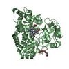
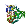
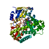
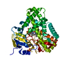
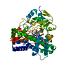
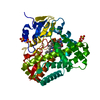
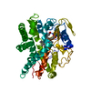
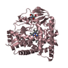
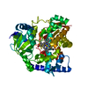
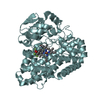
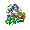
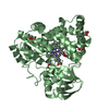
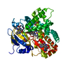
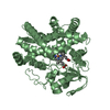
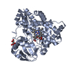
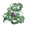
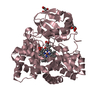
- PDB-6hqd: Cytochrome P450-153 from Pseudomonas sp. 19-rlim -

+
Open data


ID or keywords:

Loading...

-
Basic information

Entry
Database: PDB / ID: 6hqd
TitleCytochrome P450-153 from Pseudomonas sp. 19-rlim
ComponentsCytochrome P450
KeywordsOXIDOREDUCTASE / cytochrome P450 / heme / biocatalysis / Cyp153 family / METAL BINDING PROTEIN
Function / homology
Function and homology information


oxidoreductase activity, acting on paired donors, with incorporation or reduction of molecular oxygen / monooxygenase activity / iron ion binding / heme binding
Similarity search - Function
Cytochrome P450, B-class / Cytochrome p450 / Cytochrome P450 / Cytochrome P450, conserved site / Cytochrome P450 cysteine heme-iron ligand signature. / Cytochrome P450 / Cytochrome P450 superfamily / Cytochrome P450 / Orthogonal Bundle / Mainly Alpha
Similarity search - Domain/homology
PROTOPORPHYRIN IX CONTAINING FE / TRIETHYLENE GLYCOL / S,R MESO-TARTARIC ACID / Cytochrome P450
Similarity search - Component
Biological speciesPseudomonas sp. 19-rlim (bacteria)
MethodX-RAY DIFFRACTION / SYNCHROTRON / MOLECULAR REPLACEMENT / Resolution: 1.8 Å
AuthorsFiorentini, F. / Mattevi, A.
CitationJournal: Biochemistry / Year: 2018
Title: The Extreme Structural Plasticity in the CYP153 Subfamily of P450s Directs Development of Designer Hydroxylases.
Authors: Fiorentini, F. / Hatzl, A.M. / Schmidt, S. / Savino, S. / Glieder, A. / Mattevi, A.
History
DepositionSep 24, 2018Deposition site: PDBE / Processing site: PDBE
Revision 1.0Dec 12, 2018Provider: repository / Type: Initial release
Revision 1.1May 1, 2019Group: Data collection / Database references / Category: citation / pdbx_database_proc
Item: _citation.journal_id_ISSN / _citation.journal_volume ..._citation.journal_id_ISSN / _citation.journal_volume / _citation.page_first / _citation.page_last
Revision 1.2Jan 24, 2024Group: Data collection / Database references ...Data collection / Database references / Derived calculations / Refinement description
Category: chem_comp_atom / chem_comp_bond ...chem_comp_atom / chem_comp_bond / database_2 / pdbx_initial_refinement_model / struct_conn
Item: _database_2.pdbx_DOI / _database_2.pdbx_database_accession ..._database_2.pdbx_DOI / _database_2.pdbx_database_accession / _struct_conn.pdbx_dist_value / _struct_conn.ptnr1_auth_asym_id / _struct_conn.ptnr1_auth_comp_id / _struct_conn.ptnr1_auth_seq_id / _struct_conn.ptnr1_label_asym_id / _struct_conn.ptnr1_label_atom_id / _struct_conn.ptnr1_label_comp_id / _struct_conn.ptnr1_label_seq_id / _struct_conn.ptnr2_auth_asym_id / _struct_conn.ptnr2_auth_comp_id / _struct_conn.ptnr2_auth_seq_id / _struct_conn.ptnr2_label_asym_id / _struct_conn.ptnr2_label_atom_id / _struct_conn.ptnr2_label_comp_id

-
Structure visualization

Structure viewerMolecule:
MolmilJmol/JSmol

Downloads & links

-
Assembly

Deposited unit
A: Cytochrome P450
B: Cytochrome P450
C: Cytochrome P450
hetero molecules


Theoretical massNumber of molelcules
Total (without water)147,81613
Polymers145,0893
Non-polymers2,72610
Water5,783321
1
A: Cytochrome P450
hetero molecules


Theoretical massNumber of molelcules
Total (without water)49,2804
Polymers48,3631
Non-polymers9173
Water181
TypeNameSymmetry operationNumber
identity operation1_555x,y,z1
2
B: Cytochrome P450
hetero molecules


Theoretical massNumber of molelcules
Total (without water)49,2224
Polymers48,3631
Non-polymers8593
Water181
TypeNameSymmetry operationNumber
identity operation1_555x,y,z1
3
C: Cytochrome P450
hetero molecules


Theoretical massNumber of molelcules
Total (without water)49,3145
Polymers48,3631
Non-polymers9514
Water181
TypeNameSymmetry operationNumber
identity operation1_555x,y,z1
Unit cell
Length a, b, c (Å)87.311, 96.642, 87.777
Angle α, β, γ (deg.)90.00, 119.18, 90.00
Int Tables number4
Space group name H-MP1211

-
Components

-
Protein , 1 types, 3 molecules ABC

#1: Protein Cytochrome P450 /


Mass: 48363.152 Da / Num. of mol.: 3
Source method: isolated from a genetically manipulated source
Source: (gene. exp.) Pseudomonas sp. 19-rlim (bacteria) / Production host: Escherichia coli (E. coli) / Strain (production host): BL21 / References: UniProt: G3LGZ6

-
Non-polymers , 5 types, 331 molecules

#2: Chemical ChemComp-HEM / PROTOPORPHYRIN IX CONTAINING FE / HEME / Heme B


Mass: 616.487 Da / Num. of mol.: 3 / Source method: obtained synthetically / Formula: C34H32FeN4O4
#3: Chemical ChemComp-SRT / S,R MESO-TARTARIC ACID


Mass: 150.087 Da / Num. of mol.: 1 / Source method: obtained synthetically / Formula: C4H6O6
#4: Chemical ChemComp-PGE / TRIETHYLENE GLYCOL / Polyethylene glycol


Mass: 150.173 Da / Num. of mol.: 3 / Source method: obtained synthetically / Formula: C6H14O4
#5: Chemical ChemComp-GOL / GLYCEROL / GLYCERIN / PROPANE-1,2,3-TRIOL / Glycerol


Mass: 92.094 Da / Num. of mol.: 3 / Source method: obtained synthetically / Formula: C3H8O3
#6: Water ChemComp-HOH / water / Water


Mass: 18.015 Da / Num. of mol.: 321 / Source method: isolated from a natural source / Formula: H2O

-
Experimental details

-
Experiment

ExperimentMethod: X-RAY DIFFRACTION / Number of used crystals: 1

-
Sample preparation

CrystalDensity Matthews: 2.23 Å3/Da / Density % sol: 44.81 %
Crystal growTemperature: 293 K / Method: vapor diffusion / pH: 7.5 / Details: 0.2 M diammonium tartrate, 20% w/v PEG3350

-
Data collection

DiffractionMean temperature: 100 K
Diffraction sourceSource: SYNCHROTRON / Site: ESRF / Beamline: ID30B / Wavelength: 0.9677 Å
DetectorType: DECTRIS PILATUS3 X 1M / Detector: PIXEL / Date: Nov 3, 2017
RadiationProtocol: SINGLE WAVELENGTH / Monochromatic (M) / Laue (L): M / Scattering type: x-ray
Radiation wavelengthWavelength: 0.9677 Å / Relative weight: 1
ReflectionResolution: 1.8→44 Å / Num. obs: 116816 / % possible obs: 99.2 % / Redundancy: 6.7 % / CC1/2: 0.999 / Rmerge(I) obs: 0.083 / Net I/σ(I): 13.6
Reflection shellResolution: 1.8→1.83 Å / Redundancy: 6.9 % / Rmerge(I) obs: 1.155 / Num. unique obs: 5829 / CC1/2: 0.544 / % possible all: 99.9

-
Processing

Software
NameVersionClassification
REFMAC5.8.0230refinement
XDSdata reduction
Aimlessdata scaling
PHASERphasing
RefinementMethod to determine structure: MOLECULAR REPLACEMENT
Starting model: 3RWL
Resolution: 1.8→44 Å / Cor.coef. Fo:Fc: 0.966 / Cor.coef. Fo:Fc free: 0.955 / SU B: 2.902 / SU ML: 0.087 / Cross valid method: THROUGHOUT / ESU R: 0.121 / ESU R Free: 0.113 / Details: HYDROGENS HAVE BEEN ADDED IN THE RIDING POSITIONS
RfactorNum. reflection% reflectionSelection details
Rfree0.20011 5690 4.9 %RANDOM
Rwork0.1698 ---
obs0.17133 111097 99.17 %-
Solvent computationIon probe radii: 0.8 Å / Shrinkage radii: 0.8 Å / VDW probe radii: 1.2 Å
Displacement parametersBiso mean: 24.423 Å2
Baniso -1Baniso -2Baniso -3
1-0.41 Å20 Å2-0.5 Å2
2--0.56 Å20 Å2
3----0.25 Å2
Refinement stepCycle: 1 / Resolution: 1.8→44 Å
ProteinNucleic acidLigandSolventTotal
Num. atoms9929 0 176 321 10426
Refine LS restraints
Refine-IDTypeDev idealDev ideal targetNumber
X-RAY DIFFRACTIONr_bond_refined_d0.0130.01410363
X-RAY DIFFRACTIONr_bond_other_d0.0010.0179129
X-RAY DIFFRACTIONr_angle_refined_deg1.5691.68314102
X-RAY DIFFRACTIONr_angle_other_deg0.9821.64621392
X-RAY DIFFRACTIONr_dihedral_angle_1_deg6.63451238
X-RAY DIFFRACTIONr_dihedral_angle_2_deg30.36920.925584
X-RAY DIFFRACTIONr_dihedral_angle_3_deg13.95151711
X-RAY DIFFRACTIONr_dihedral_angle_4_deg16.4651595
X-RAY DIFFRACTIONr_chiral_restr0.0780.21320
X-RAY DIFFRACTIONr_gen_planes_refined0.0080.0211677
X-RAY DIFFRACTIONr_gen_planes_other0.0030.021927
X-RAY DIFFRACTIONr_nbd_refined
X-RAY DIFFRACTIONr_nbd_other
X-RAY DIFFRACTIONr_nbtor_refined
X-RAY DIFFRACTIONr_nbtor_other
X-RAY DIFFRACTIONr_xyhbond_nbd_refined
X-RAY DIFFRACTIONr_xyhbond_nbd_other
X-RAY DIFFRACTIONr_metal_ion_refined
X-RAY DIFFRACTIONr_metal_ion_other
X-RAY DIFFRACTIONr_symmetry_vdw_refined
X-RAY DIFFRACTIONr_symmetry_vdw_other
X-RAY DIFFRACTIONr_symmetry_hbond_refined
X-RAY DIFFRACTIONr_symmetry_hbond_other
X-RAY DIFFRACTIONr_symmetry_metal_ion_refined
X-RAY DIFFRACTIONr_symmetry_metal_ion_other
X-RAY DIFFRACTIONr_mcbond_it1.8782.3374961
X-RAY DIFFRACTIONr_mcbond_other1.8742.3364960
X-RAY DIFFRACTIONr_mcangle_it2.5133.4926196
X-RAY DIFFRACTIONr_mcangle_other2.5133.4926197
X-RAY DIFFRACTIONr_scbond_it3.0552.6865402
X-RAY DIFFRACTIONr_scbond_other3.0552.6865402
X-RAY DIFFRACTIONr_scangle_it
X-RAY DIFFRACTIONr_scangle_other4.5763.8897907
X-RAY DIFFRACTIONr_long_range_B_refined5.33526.95111142
X-RAY DIFFRACTIONr_long_range_B_other5.33826.93411121
X-RAY DIFFRACTIONr_rigid_bond_restr
X-RAY DIFFRACTIONr_sphericity_free
X-RAY DIFFRACTIONr_sphericity_bonded
LS refinement shellResolution: 1.8→1.847 Å / Total num. of bins used: 20
RfactorNum. reflection% reflection
Rfree0.3 498 -
Rwork0.282 8217 -
obs--99.89 %

+
About Yorodumi

-
News

-
Feb 9, 2022. New format data for meta-information of EMDB entries

New format data for meta-information of EMDB entries

  • Version 3 of the EMDB header file is now the official format.
  • The previous official version 1.9 will be removed from the archive.

Related info.:EMDB header

External links:wwPDB to switch to version 3 of the EMDB data model

-
Aug 12, 2020. Covid-19 info

Covid-19 info

URL: https://pdbj.org/emnavi/covid19.php

New page: Covid-19 featured information page in EM Navigator.

Related info.:Covid-19 info / Mar 5, 2020. Novel coronavirus structure data

+
Mar 5, 2020. Novel coronavirus structure data

Novel coronavirus structure data

Related info.:Yorodumi Speices / Aug 12, 2020. Covid-19 info

External links:COVID-19 featured content - PDBj / Molecule of the Month (242):Coronavirus Proteases

+
Jan 31, 2019. EMDB accession codes are about to change! (news from PDBe EMDB page)

EMDB accession codes are about to change! (news from PDBe EMDB page)

  • The allocation of 4 digits for EMDB accession codes will soon come to an end. Whilst these codes will remain in use, new EMDB accession codes will include an additional digit and will expand incrementally as the available range of codes is exhausted. The current 4-digit format prefixed with “EMD-” (i.e. EMD-XXXX) will advance to a 5-digit format (i.e. EMD-XXXXX), and so on. It is currently estimated that the 4-digit codes will be depleted around Spring 2019, at which point the 5-digit format will come into force.
  • The EM Navigator/Yorodumi systems omit the EMD- prefix.

Related info.:Q: What is EMD? / ID/Accession-code notation in Yorodumi/EM Navigator

External links:EMDB Accession Codes are Changing Soon! / Contact to PDBj

+
Jul 12, 2017. Major update of PDB

Major update of PDB

  • wwPDB released updated PDB data conforming to the new PDBx/mmCIF dictionary.
  • This is a major update changing the version number from 4 to 5, and with Remediation, in which all the entries are updated.
  • In this update, many items about electron microscopy experimental information are reorganized (e.g. em_software).
  • Now, EM Navigator and Yorodumi are based on the updated data.

External links:wwPDB Remediation / Enriched Model Files Conforming to OneDep Data Standards Now Available in the PDB FTP Archive

-
Yorodumi

Thousand views of thousand structures

  • Yorodumi is a browser for structure data from EMDB, PDB, SASBDB, etc.
  • This page is also the successor to EM Navigator detail page, and also detail information page/front-end page for Omokage search.
  • The word "yorodu" (or yorozu) is an old Japanese word meaning "ten thousand". "mi" (miru) is to see.

Related info.:EMDB / PDB / SASBDB / Comparison of 3 databanks / Yorodumi Search / Aug 31, 2016. New EM Navigator & Yorodumi / Yorodumi Papers / Jmol/JSmol / Function and homology information / Changes in new EM Navigator and Yorodumi

Read more