 Movie
Movie Controller

[English] 日本語
 Yorodumi
- PDB-1lw1: Crystal Structure Of Mycobacterium Tuberculosis Alkylperoxidase A... -
+ Open data

- Basic information
| Entry | Database: PDB / ID: 1lw1 | ||||||
|---|---|---|---|---|---|---|---|
| Title | Crystal Structure Of Mycobacterium Tuberculosis Alkylperoxidase Ahpd H137F mutant | ||||||
 Components Components | ALKYLHYDROPEROXIDASE D | ||||||
 Keywords Keywords | OXIDOREDUCTASE / ALKYLHYDROPEROXIDASE / TUBERCULOSIS | ||||||
| Function / homology |  Function and homology information Function and homology informationlipoyl-dependent peroxiredoxin / : / : / Cell redox homeostasis / disulfide oxidoreductase activity / peroxiredoxin activity / cell redox homeostasis / peroxidase activity / response to oxidative stress / oxidoreductase activity ...lipoyl-dependent peroxiredoxin / : / : / Cell redox homeostasis / disulfide oxidoreductase activity / peroxiredoxin activity / cell redox homeostasis / peroxidase activity / response to oxidative stress / oxidoreductase activity / plasma membrane / cytosol Similarity search - Function | ||||||
| Biological species |   Mycobacterium tuberculosis (bacteria) Mycobacterium tuberculosis (bacteria) | ||||||
| Method |  X-RAY DIFFRACTION / SYNCHROTRON / MOLECULAR REPLACEMENT / Resolution: 2.3 Å X-RAY DIFFRACTION / SYNCHROTRON / MOLECULAR REPLACEMENT / Resolution: 2.3 Å | ||||||
 Authors Authors | Nunn, C.M. / Djordjevic, S. / Ortiz de Montellano, P.R. | ||||||
 Citation Citation | Journal: J.Biol.Chem. / Year: 2003 Title: The Mechanism of Mycobacterium tuberculosis Alkylhydroperoxidase AhpD as Defined by Mutagenesis, Crystallography, and Kinetics Authors: Koshkin, A. / Nunn, C.M. / Djordjevic, S. / Ortiz de Montellano, P.R. #1: Journal: Acta Crystallogr.,Sect.D / Year: 1998Title: Crystallography & NMR System: A New Software Suite for Macromolecular Structure Determination Authors: Brunger, A.T. / Adams, P.D. / Clore, G.M. / Delano, W.L. / Gros, P. / Grosse-Kunstleve, R.W. / Jiang, J.-S. / Kuszewski, J. / Nilges, M. / Pannu, N.S. / Read, R.J. / Rice, L.M. / Simonson, T. / Warren, G.L. | ||||||
| History |
|
- Structure visualization
| Structure viewer | Molecule: MolmilJmol/JSmol |
|---|
- Downloads & links
-Download
| PDBx/mmCIF format | 1lw1.cif.gz | 114.5 KB | Display | PDBx/mmCIF format |
|---|---|---|---|---|
| PDB format | pdb1lw1.ent.gz | 89.7 KB | Display | PDB format |
| PDBx/mmJSON format | 1lw1.json.gz | Tree view | PDBx/mmJSON format | |
| Others |  Other downloads Other downloads |
-Validation report
| Arichive directory | https://data.pdbj.org/pub/pdb/validation_reports/lw/1lw1ftp://data.pdbj.org/pub/pdb/validation_reports/lw/1lw1 | HTTPS FTP |
|---|
-Related structure data
| Related structure data | 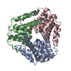 1me5C  1gu9S S: Starting model for refinement C: citing same article ( |
|---|---|
| Similar structure data |

-Links
 PDBj
PDBj- Assembly
| Deposited unit | 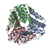
| ||||||||||||
|---|---|---|---|---|---|---|---|---|---|---|---|---|---|
| 1 |
| ||||||||||||
| 2 | 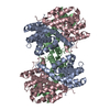
| ||||||||||||
| Unit cell |
| ||||||||||||
| Components on special symmetry positions |
| ||||||||||||
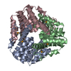
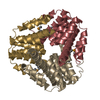
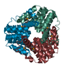
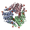
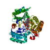
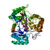
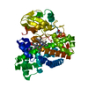
| Details | Chains A, B and C form a protein trimer |
-Components
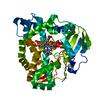
| #1: Protein | Mass: 18810.547 Da / Num. of mol.: 3 / Mutation: h137f Source method: isolated from a genetically manipulated source Source: (gene. exp.) Mycobacterium tuberculosis (bacteria) / Strain: H37Rv / Plasmid: pACAhpD / Species (production host): Escherichia coli / Production host: #2: Water | ChemComp-HOH / |  Mass: 18.015 Da / Num. of mol.: 364 / Source method: isolated from a natural source / Formula: H2O Mass: 18.015 Da / Num. of mol.: 364 / Source method: isolated from a natural source / Formula: H2O |
|---|
-Experimental details
-Experiment
| Experiment | Method: X-RAY DIFFRACTION / Number of used crystals: 1 |
|---|
- Sample preparation
| Crystal | Density Matthews: 1.96 Å3/Da / Density % sol: 37 % | ||||||||||||||||||||||||||||||||||||||||||||||||||||||||||||||||||||||
|---|---|---|---|---|---|---|---|---|---|---|---|---|---|---|---|---|---|---|---|---|---|---|---|---|---|---|---|---|---|---|---|---|---|---|---|---|---|---|---|---|---|---|---|---|---|---|---|---|---|---|---|---|---|---|---|---|---|---|---|---|---|---|---|---|---|---|---|---|---|---|---|
| Crystal grow | Temperature: 290 K / Method: vapor diffusion, hanging drop / pH: 6 Details: 100MM SODIUM CITRATE BUFFER, PH 5.6, CONTAINING 200MM AMMONIUM ACETATE AND 26% PEG 4000. MIXED IN EQUAL VOLUME WITH AHPD (4.5 MG/ML) IN 25MM MOPS BUFFER, PH 7.2, CONTAINING 50MM KCL, 10% ...Details: 100MM SODIUM CITRATE BUFFER, PH 5.6, CONTAINING 200MM AMMONIUM ACETATE AND 26% PEG 4000. MIXED IN EQUAL VOLUME WITH AHPD (4.5 MG/ML) IN 25MM MOPS BUFFER, PH 7.2, CONTAINING 50MM KCL, 10% GLYCEROL, 0.1MM EDTA, VAPOR DIFFUSION, HANGING DROP, temperature 290.0K | ||||||||||||||||||||||||||||||||||||||||||||||||||||||||||||||||||||||
| Crystal grow | *PLUS pH: 7.2 | ||||||||||||||||||||||||||||||||||||||||||||||||||||||||||||||||||||||
| Components of the solutions | *PLUS
|
-Data collection
| Diffraction | Mean temperature: 110 K |
|---|---|
| Diffraction source | Source: SYNCHROTRON / Site: SRS  / Beamline: PX9.6 / Wavelength: 0.87 Å / Beamline: PX9.6 / Wavelength: 0.87 Å |
| Detector | Type: ADSC QUANTUM 4 / Detector: CCD / Date: Apr 16, 2002 / Details: mirrors |
| Radiation | Monochromator: Single crystal Si(111) / Protocol: SINGLE WAVELENGTH / Monochromatic (M) / Laue (L): M / Scattering type: x-ray |
| Radiation wavelength | Wavelength: 0.87 Å / Relative weight: 1 |
| Reflection | Resolution: 2.3→50 Å / Num. all: 18648 / Num. obs: 18396 / % possible obs: 92.5 % / Observed criterion σ(F): 2 / Observed criterion σ(I): 2 / Redundancy: 3.42 % / Biso Wilson estimate: 32.6 Å2 / Rmerge(I) obs: 0.11 / Net I/σ(I): 9.188 |
| Reflection shell | Resolution: 2.3→2.38 Å / Redundancy: 3 % / Rmerge(I) obs: 0.24 / Mean I/σ(I) obs: 2.94 / % possible all: 70.3 |
| Reflection | *PLUS Lowest resolution: 50 Å / Num. measured all: 396017 |
| Reflection shell | *PLUS % possible obs: 70.3 % |
- Processing
| Software |
| ||||||||||||||||||||||||||||||||||||
|---|---|---|---|---|---|---|---|---|---|---|---|---|---|---|---|---|---|---|---|---|---|---|---|---|---|---|---|---|---|---|---|---|---|---|---|---|---|
| Refinement | Method to determine structure: MOLECULAR REPLACEMENT Starting model: 1GU9 Resolution: 2.3→50 Å / Isotropic thermal model: restrained / Cross valid method: THROUGHOUT / σ(F): 2 / Stereochemistry target values: Engh & Huber
| ||||||||||||||||||||||||||||||||||||
| Solvent computation | Solvent model: CNS bulk solvent model used / Bsol: 75.3875 Å2 / ksol: 0.352637 e/Å3 | ||||||||||||||||||||||||||||||||||||
| Displacement parameters | Biso mean: 44.14 Å2
| ||||||||||||||||||||||||||||||||||||
| Refine analyze |
| ||||||||||||||||||||||||||||||||||||
| Refinement step | Cycle: LAST / Resolution: 2.3→50 Å
| ||||||||||||||||||||||||||||||||||||
| Refine LS restraints |
| ||||||||||||||||||||||||||||||||||||
| LS refinement shell | Resolution: 2.3→2.38 Å / Rfactor Rfree error: 0.006 / Total num. of bins used: 10
| ||||||||||||||||||||||||||||||||||||
| Xplor file |
| ||||||||||||||||||||||||||||||||||||
| Refinement | *PLUS Lowest resolution: 50 Å / Rfactor Rwork: 0.184 | ||||||||||||||||||||||||||||||||||||
| Solvent computation | *PLUS | ||||||||||||||||||||||||||||||||||||
| Displacement parameters | *PLUS | ||||||||||||||||||||||||||||||||||||
| Refine LS restraints | *PLUS
|
