 Movie
Movie Controller


+ Open data

- Basic information
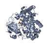
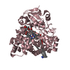
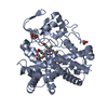
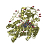
| Entry | Database: PDB / ID: 6l39 | ||||||
|---|---|---|---|---|---|---|---|
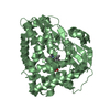
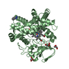
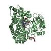
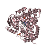
| Title | Cytochrome P450 107G1 (RapN) | ||||||
 Components Components | Cytochrome P450 | ||||||
 Keywords Keywords | OXIDOREDUCTASE / Cytochrome P450 | ||||||
| Function / homology |  Function and homology information Function and homology informationoxidoreductase activity, acting on paired donors, with incorporation or reduction of molecular oxygen / monooxygenase activity / iron ion binding / heme binding Similarity search - Function | ||||||
| Biological species | Streptomyces rapamycinicus | ||||||
| Method |  X-RAY DIFFRACTION / SYNCHROTRON / MOLECULAR REPLACEMENT / Resolution: 2.97 Å X-RAY DIFFRACTION / SYNCHROTRON / MOLECULAR REPLACEMENT / Resolution: 2.97 Å | ||||||
 Authors Authors | Kim, V.C. / Kim, D.H. / Lim, Y.R. / Lee, I.H. / Lee, J.H. / Kang, L.W. | ||||||
 Citation Citation | Journal: Arch.Biochem.Biophys. / Year: 2020 Title: Structural insights into CYP107G1 from rapamycin-producing Streptomyces rapamycinicus. Authors: Kim, V. / Lim, Y.R. / Lee, I. / Lee, J.H. / Han, S. / Pham, T.V. / Kim, H. / Lee, R. / Kang, L.W. / Kim, D. | ||||||
| History |
|
- Structure visualization
| Structure viewer | Molecule: MolmilJmol/JSmol |
|---|
- Downloads & links
-Download
| PDBx/mmCIF format | 6l39.cif.gz | 161.9 KB | Display | PDBx/mmCIF format |
|---|---|---|---|---|
| PDB format | pdb6l39.ent.gz | 126.1 KB | Display | PDB format |
| PDBx/mmJSON format | 6l39.json.gz | Tree view | PDBx/mmJSON format | |
| Others |  Other downloads Other downloads |
-Validation report
| Arichive directory | https://data.pdbj.org/pub/pdb/validation_reports/l3/6l39ftp://data.pdbj.org/pub/pdb/validation_reports/l3/6l39 | HTTPS FTP |
|---|
-Related structure data
| Related structure data |  6l3aC  4z5pS S: Starting model for refinement C: citing same article ( |
|---|---|
| Similar structure data |


-Links
 PDBj
PDBj
- Assembly
| Deposited unit | 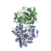
| ||||||||
|---|---|---|---|---|---|---|---|---|---|
| 1 | 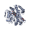
| ||||||||
| 2 | 
| ||||||||
| Unit cell |
|
-Components
| #1: Protein | Mass: 45134.879 Da / Num. of mol.: 2 Source method: isolated from a genetically manipulated source Source: (gene. exp.)  Streptomyces rapamycinicus (strain ATCC 29253 / DSM 41530 / NRRL 5491 / AYB-994) (bacteria) Streptomyces rapamycinicus (strain ATCC 29253 / DSM 41530 / NRRL 5491 / AYB-994) (bacteria)Strain: ATCC 29253 / DSM 41530 / NRRL 5491 / AYB-994 / Gene: D3C57_102975 / Plasmid: pET28a+ / Production host: #2: Chemical |   Mass: 616.487 Da / Num. of mol.: 2 / Source method: obtained synthetically / Formula: C34H32FeN4O4 / Feature type: SUBJECT OF INVESTIGATION Mass: 616.487 Da / Num. of mol.: 2 / Source method: obtained synthetically / Formula: C34H32FeN4O4 / Feature type: SUBJECT OF INVESTIGATION#3: Chemical |   Mass: 106.120 Da / Num. of mol.: 2 / Source method: obtained synthetically / Formula: C4H10O3 Mass: 106.120 Da / Num. of mol.: 2 / Source method: obtained synthetically / Formula: C4H10O3#4: Chemical |   Mass: 94.971 Da / Num. of mol.: 2 / Source method: obtained synthetically / Formula: PO4 Mass: 94.971 Da / Num. of mol.: 2 / Source method: obtained synthetically / Formula: PO4#5: Water | ChemComp-HOH / |  Mass: 18.015 Da / Num. of mol.: 4 / Source method: isolated from a natural source / Formula: H2O Mass: 18.015 Da / Num. of mol.: 4 / Source method: isolated from a natural source / Formula: H2OHas ligand of interest | Y | |
|---|
-Experimental details
-Experiment
| Experiment | Method: X-RAY DIFFRACTION / Number of used crystals: 1 |
|---|
- Sample preparation
| Crystal | Density Matthews: 2.08 Å3/Da / Density % sol: 40.82 % |
|---|---|
| Crystal grow | Temperature: 287.15 K / Method: vapor diffusion, sitting drop / pH: 8 / Details: 0.1 M imidazole pH 8.0 1 M sodium citrate |
-Data collection
| Diffraction | Mean temperature: 100 K / Serial crystal experiment: N |
|---|---|
| Diffraction source | Source: SYNCHROTRON / Site: PAL/PLS  / Beamline: 5C (4A) / Wavelength: 0.9794 Å / Beamline: 5C (4A) / Wavelength: 0.9794 Å |
| Detector | Type: ADSC QUANTUM 315r / Detector: CCD / Date: Dec 14, 2016 |
| Radiation | Protocol: SINGLE WAVELENGTH / Monochromatic (M) / Laue (L): M / Scattering type: x-ray |
| Radiation wavelength | Wavelength: 0.9794 Å / Relative weight: 1 |
| Reflection | Resolution: 2.9→50 Å / Num. obs: 39493 / % possible obs: 96.6 % / Redundancy: 2.8 % / CC1/2: 0.949 / Net I/σ(I): 8.6 |
| Reflection shell | Resolution: 2.9→2.95 Å / Mean I/σ(I) obs: 8.6 / Num. unique obs: 39493 / CC1/2: 0.949 |
- Processing
| Software |
| ||||||||||||||||||||||||
|---|---|---|---|---|---|---|---|---|---|---|---|---|---|---|---|---|---|---|---|---|---|---|---|---|---|
| Refinement | Method to determine structure: MOLECULAR REPLACEMENT Starting model: 4Z5P Resolution: 2.97→32.77 Å / Cor.coef. Fo:Fc: 0.95 / Cor.coef. Fo:Fc free: 0.837 / SU B: 0.017 / SU ML: 0 / Cross valid method: THROUGHOUT / σ(F): 0 / ESU R: 0.398 / ESU R Free: 0.63 / Stereochemistry target values: MAXIMUM LIKELIHOOD Details: HYDROGENS HAVE BEEN ADDED IN THE RIDING POSITIONS U VALUES : REFINED INDIVIDUALLY
| ||||||||||||||||||||||||
| Solvent computation | Ion probe radii: 0.8 Å / Shrinkage radii: 0.8 Å / VDW probe radii: 1.2 Å / Solvent model: MASK | ||||||||||||||||||||||||
| Displacement parameters | Biso max: 140.65 Å2 / Biso mean: 69.884 Å2 / Biso min: 30 Å2
| ||||||||||||||||||||||||
| Refinement step | Cycle: final / Resolution: 2.97→32.77 Å
| ||||||||||||||||||||||||
| LS refinement shell | Resolution: 2.972→3.049 Å / Rfactor Rfree error: 0 / Total num. of bins used: 20
|
