 Movie
Movie Controller


+ Open data

- Basic information
| Entry | Database: PDB / ID: 6zi3 | ||||||
|---|---|---|---|---|---|---|---|
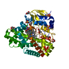
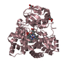
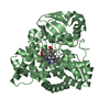
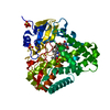
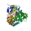
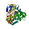
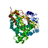
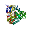
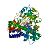
| Title | Crystal structure of OleP-6DEB bound to L-rhamnose | ||||||
 Components Components | Cytochrome P-450 | ||||||
 Keywords Keywords | OXIDOREDUCTASE / cytochrome P450 / 8.8a-deoxyoleandolide monooxygenase / 8.8a-deoxyoleandolide | ||||||
| Function / homology |  Function and homology information Function and homology informationoxidoreductase activity, acting on paired donors, with incorporation or reduction of molecular oxygen / monooxygenase activity / iron ion binding / heme binding Similarity search - Function | ||||||
| Biological species |  Streptomyces antibioticus (bacteria) Streptomyces antibioticus (bacteria) | ||||||
| Method |  X-RAY DIFFRACTION / SYNCHROTRON / FOURIER SYNTHESIS / Resolution: 2.08 Å X-RAY DIFFRACTION / SYNCHROTRON / FOURIER SYNTHESIS / Resolution: 2.08 Å | ||||||
 Authors Authors | Montemiglio, L.C. / Savino, C. / Vallone, B. / Parisi, G. / Freda, I. | ||||||
| Funding support |  Italy, 1items Italy, 1items
| ||||||
 Citation Citation | Journal: Biomolecules / Year: 2020 Title: Dissecting the Cytochrome P450 OleP Substrate Specificity: Evidence for a Preferential Substrate. Authors: Parisi, G. / Freda, I. / Exertier, C. / Cecchetti, C. / Gugole, E. / Cerutti, G. / D'Auria, L. / Macone, A. / Vallone, B. / Savino, C. / Montemiglio, L.C. | ||||||
| History |
|
- Structure visualization
| Structure viewer | Molecule: MolmilJmol/JSmol |
|---|
- Downloads & links
-Download
| PDBx/mmCIF format | 6zi3.cif.gz | 1 MB | Display | PDBx/mmCIF format |
|---|---|---|---|---|
| PDB format | pdb6zi3.ent.gz | 897.7 KB | Display | PDB format |
| PDBx/mmJSON format | 6zi3.json.gz | Tree view | PDBx/mmJSON format | |
| Others |  Other downloads Other downloads |
-Validation report
| Arichive directory | https://data.pdbj.org/pub/pdb/validation_reports/zi/6zi3ftp://data.pdbj.org/pub/pdb/validation_reports/zi/6zi3 | HTTPS FTP |
|---|
-Related structure data
| Related structure data |  6zhzC  6zi2C  6zi7C  5mnsS C: citing same article ( S: Starting model for refinement |
|---|---|
| Similar structure data |

-Links
 PDBj
PDBj
- Assembly
| Deposited unit | 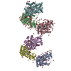
| ||||||||
|---|---|---|---|---|---|---|---|---|---|
| 1 | 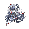
| ||||||||
| 2 | 
| ||||||||
| 3 | 
| ||||||||
| 4 | 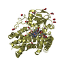
| ||||||||
| 5 | 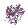
| ||||||||
| 6 | 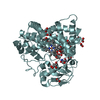
| ||||||||
| Unit cell |
|
-Components
-Protein / Sugars , 2 types, 13 molecules ABCDEF
| #1: Protein | Mass: 45012.070 Da / Num. of mol.: 6 Source method: isolated from a genetically manipulated source Source: (gene. exp.) Streptomyces antibioticus (bacteria) / Gene: oleP / Production host: #4: Sugar | ChemComp-RAM /  Type: L-saccharide, alpha linking / Mass: 164.156 Da / Num. of mol.: 7 Type: L-saccharide, alpha linking / Mass: 164.156 Da / Num. of mol.: 7Source method: isolated from a genetically manipulated source Formula: C6H12O5 |
|---|
-Non-polymers , 7 types, 1390 molecules 


| #2: Chemical | ChemComp-HEM /  Mass: 616.487 Da / Num. of mol.: 6 / Source method: obtained synthetically / Formula: C34H32FeN4O4 Mass: 616.487 Da / Num. of mol.: 6 / Source method: obtained synthetically / Formula: C34H32FeN4O4#3: Chemical | ChemComp-DEB /  Mass: 386.523 Da / Num. of mol.: 6 / Source method: obtained synthetically / Formula: C21H38O6 Mass: 386.523 Da / Num. of mol.: 6 / Source method: obtained synthetically / Formula: C21H38O6#5: Chemical |  Mass: 122.143 Da / Num. of mol.: 3 / Source method: obtained synthetically / Formula: C4H12NO3 / Comment: pH buffer*YM Mass: 122.143 Da / Num. of mol.: 3 / Source method: obtained synthetically / Formula: C4H12NO3 / Comment: pH buffer*YM#6: Chemical | ChemComp-FMT /  Mass: 46.025 Da / Num. of mol.: 203 / Source method: obtained synthetically / Formula: CH2O2 Mass: 46.025 Da / Num. of mol.: 203 / Source method: obtained synthetically / Formula: CH2O2#7: Chemical | ChemComp-NA /  Mass: 22.990 Da / Num. of mol.: 7 / Source method: obtained synthetically / Formula: Na Mass: 22.990 Da / Num. of mol.: 7 / Source method: obtained synthetically / Formula: Na#8: Chemical | ChemComp-GOL /  Mass: 92.094 Da / Num. of mol.: 6 / Source method: obtained synthetically / Formula: C3H8O3 Mass: 92.094 Da / Num. of mol.: 6 / Source method: obtained synthetically / Formula: C3H8O3#9: Water | ChemComp-HOH / | Mass: 18.015 Da / Num. of mol.: 1159 / Source method: isolated from a natural source / Formula: H2O |
|---|
-Details
| Has ligand of interest | N |
|---|
-Experimental details
-Experiment
| Experiment | Method: X-RAY DIFFRACTION / Number of used crystals: 1 |
|---|
- Sample preparation
| Crystal | Density Matthews: 3.1 Å3/Da / Density % sol: 60.5 % |
|---|---|
| Crystal grow | Temperature: 294 K / Method: vapor diffusion, hanging drop / Details: 4.2 M Sodium Formate (HCOONa) |
-Data collection
| Diffraction | Mean temperature: 100 K / Serial crystal experiment: N |
|---|---|
| Diffraction source | Source: SYNCHROTRON / Site: ELETTRA / Beamline: 11.2C / Wavelength: 1 Å |
| Detector | Type: DECTRIS PILATUS 6M / Detector: PIXEL / Date: May 16, 2019 |
| Radiation | Protocol: SINGLE WAVELENGTH / Monochromatic (M) / Laue (L): M / Scattering type: x-ray |
| Radiation wavelength | Wavelength: 1 Å / Relative weight: 1 |
| Reflection | Resolution: 1.96→50 Å / Num. obs: 462663 / % possible obs: 97.7 % / Redundancy: 2.7 % / CC1/2: 0.997 / Rmerge(I) obs: 0.064 / Net I/σ(I): 7.8 |
| Reflection shell | Resolution: 1.96→2.08 Å / Rmerge(I) obs: 1.64 / Num. unique obs: 73872 / CC1/2: 0.362 |
- Processing
| Software |
| ||||||||||||||||||||||||||||||||||||||||||||||||||||||||||||||||||||||||||||||||||||||||||||||||||||||||||||||||||||||||||||||||||||||||||||||||||||||||||||||||||||||||||||||||||||||
|---|---|---|---|---|---|---|---|---|---|---|---|---|---|---|---|---|---|---|---|---|---|---|---|---|---|---|---|---|---|---|---|---|---|---|---|---|---|---|---|---|---|---|---|---|---|---|---|---|---|---|---|---|---|---|---|---|---|---|---|---|---|---|---|---|---|---|---|---|---|---|---|---|---|---|---|---|---|---|---|---|---|---|---|---|---|---|---|---|---|---|---|---|---|---|---|---|---|---|---|---|---|---|---|---|---|---|---|---|---|---|---|---|---|---|---|---|---|---|---|---|---|---|---|---|---|---|---|---|---|---|---|---|---|---|---|---|---|---|---|---|---|---|---|---|---|---|---|---|---|---|---|---|---|---|---|---|---|---|---|---|---|---|---|---|---|---|---|---|---|---|---|---|---|---|---|---|---|---|---|---|---|---|---|
| Refinement | Method to determine structure: FOURIER SYNTHESIS Starting model: 5MNS Resolution: 2.08→48.09 Å / Cor.coef. Fo:Fc: 0.974 / Cor.coef. Fo:Fc free: 0.952 / SU B: 10.753 / SU ML: 0.137 / Cross valid method: THROUGHOUT / ESU R: 0.174 / ESU R Free: 0.165 / Stereochemistry target values: MAXIMUM LIKELIHOOD / Details: HYDROGENS HAVE BEEN ADDED IN THE RIDING POSITIONS
| ||||||||||||||||||||||||||||||||||||||||||||||||||||||||||||||||||||||||||||||||||||||||||||||||||||||||||||||||||||||||||||||||||||||||||||||||||||||||||||||||||||||||||||||||||||||
| Solvent computation | Ion probe radii: 0.8 Å / Shrinkage radii: 0.8 Å / VDW probe radii: 1.2 Å / Solvent model: MASK | ||||||||||||||||||||||||||||||||||||||||||||||||||||||||||||||||||||||||||||||||||||||||||||||||||||||||||||||||||||||||||||||||||||||||||||||||||||||||||||||||||||||||||||||||||||||
| Displacement parameters | Biso mean: 56.045 Å2
| ||||||||||||||||||||||||||||||||||||||||||||||||||||||||||||||||||||||||||||||||||||||||||||||||||||||||||||||||||||||||||||||||||||||||||||||||||||||||||||||||||||||||||||||||||||||
| Refinement step | Cycle: 1 / Resolution: 2.08→48.09 Å
| ||||||||||||||||||||||||||||||||||||||||||||||||||||||||||||||||||||||||||||||||||||||||||||||||||||||||||||||||||||||||||||||||||||||||||||||||||||||||||||||||||||||||||||||||||||||
| Refine LS restraints |
|