 Movie
Movie Controller

[English] 日本語
 Yorodumi
- PDB-4iq7: Substrate and reaction specificity of Mycobacterium tuberculosis ... -
+ Open data

- Basic information
| Entry | Database: PDB / ID: 4iq7 | ||||||
|---|---|---|---|---|---|---|---|
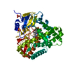
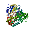
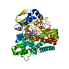
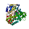
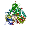
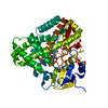
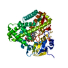
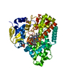
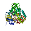
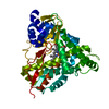
| Title | Substrate and reaction specificity of Mycobacterium tuberculosis cytochrome P450 CYP121 | ||||||
 Components Components | Cytochrome P450 121 | ||||||
 Keywords Keywords | OXIDOREDUCTASE / protein-ligand complex / P450 fold / oxidase | ||||||
| Function / homology |  Function and homology information Function and homology informationmycocyclosin synthase / oxidoreductase activity, acting on paired donors, with oxidation of a pair of donors resulting in the reduction of molecular oxygen to two molecules of water / cyclase activity / carbon monoxide binding / monooxygenase activity / oxidoreductase activity / iron ion binding / heme binding / cytoplasm Similarity search - Function | ||||||
| Biological species |   Mycobacterium tuberculosis (bacteria) Mycobacterium tuberculosis (bacteria) | ||||||
| Method |  X-RAY DIFFRACTION / SYNCHROTRON / FOURIER SYNTHESIS / Resolution: 1.9 Å X-RAY DIFFRACTION / SYNCHROTRON / FOURIER SYNTHESIS / Resolution: 1.9 Å | ||||||
 Authors Authors | Fonvielle, M. / Ledu, M.H. / Lequin, O. / Lecoq, A. / Jacquet, M. / Thai, R. / Dubois, S. / Grach, G. / Gondry, M. / Belin, P. | ||||||
 Citation Citation | Journal: J.Biol.Chem. / Year: 2013 Title: Substrate and Reaction Specificity of Mycobacterium tuberculosis Cytochrome P450 CYP121: INSIGHTS FROM BIOCHEMICAL STUDIES AND CRYSTAL STRUCTURES. Authors: Fonvielle, M. / Le Du, M.H. / Lequin, O. / Lecoq, A. / Jacquet, M. / Thai, R. / Dubois, S. / Grach, G. / Gondry, M. / Belin, P. | ||||||
| History |
|
- Structure visualization
| Structure viewer | Molecule: MolmilJmol/JSmol |
|---|
- Downloads & links
-Download
| PDBx/mmCIF format | 4iq7.cif.gz | 181.4 KB | Display | PDBx/mmCIF format |
|---|---|---|---|---|
| PDB format | pdb4iq7.ent.gz | 141.5 KB | Display | PDB format |
| PDBx/mmJSON format | 4iq7.json.gz | Tree view | PDBx/mmJSON format | |
| Others |  Other downloads Other downloads |
-Validation report
| Arichive directory | https://data.pdbj.org/pub/pdb/validation_reports/iq/4iq7ftp://data.pdbj.org/pub/pdb/validation_reports/iq/4iq7 | HTTPS FTP |
|---|
-Related structure data


-Links
 PDBj
PDBj
- Assembly
| Deposited unit | 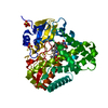
| ||||||||||||
|---|---|---|---|---|---|---|---|---|---|---|---|---|---|
| 1 |
| ||||||||||||
| 2 | 
| ||||||||||||
| 3 | 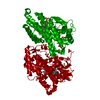
| ||||||||||||
| Unit cell |
| ||||||||||||
| Components on special symmetry positions |
|
-Components
| #1: Protein | Mass: 43174.668 Da / Num. of mol.: 1 Source method: isolated from a genetically manipulated source Source: (gene. exp.) Mycobacterium tuberculosis (bacteria) / Gene: cyp121, Rv2276, MT2336, MTCY339.34c / Production host: References: UniProt: P0A514, UniProt: P9WPP7*PLUS, Oxidoreductases; Acting on paired donors, with incorporation or reduction of molecular oxygen | ||||
|---|---|---|---|---|---|
| #2: Chemical | ChemComp-HEM /   Mass: 616.487 Da / Num. of mol.: 1 / Source method: obtained synthetically / Formula: C34H32FeN4O4 Mass: 616.487 Da / Num. of mol.: 1 / Source method: obtained synthetically / Formula: C34H32FeN4O4 | ||||
| #3: Chemical | ChemComp-SO4 /   Mass: 96.063 Da / Num. of mol.: 5 / Source method: obtained synthetically / Formula: SO4 Mass: 96.063 Da / Num. of mol.: 5 / Source method: obtained synthetically / Formula: SO4#4: Chemical | ChemComp-1G9 / ( |   Mass: 234.251 Da / Num. of mol.: 1 / Source method: obtained synthetically / Formula: C12H14N2O3 Mass: 234.251 Da / Num. of mol.: 1 / Source method: obtained synthetically / Formula: C12H14N2O3#5: Water | ChemComp-HOH / |  Mass: 18.015 Da / Num. of mol.: 605 / Source method: isolated from a natural source / Formula: H2O Mass: 18.015 Da / Num. of mol.: 605 / Source method: isolated from a natural source / Formula: H2O |
-Experimental details
-Experiment
| Experiment | Method: X-RAY DIFFRACTION / Number of used crystals: 1 |
|---|
- Sample preparation
| Crystal | Density Matthews: 2.68 Å3/Da / Density % sol: 54.05 % |
|---|---|
| Crystal grow | Temperature: 277 K / Method: vapor diffusion, sitting drop / pH: 5 Details: ammonium sulfate 2.2 M NaMes 0.1 M, pH 5, VAPOR DIFFUSION, SITTING DROP, temperature 277K |
-Data collection
| Diffraction | Mean temperature: 100 K |
|---|---|
| Diffraction source | Source: SYNCHROTRON / Site: ESRF  / Beamline: ID23-1 / Wavelength: 0.933 Å / Beamline: ID23-1 / Wavelength: 0.933 Å |
| Detector | Type: ADSC QUANTUM 315 / Detector: CCD / Date: Jul 31, 2010 |
| Radiation | Monochromator: monochromator crystal / Protocol: SINGLE WAVELENGTH / Monochromatic (M) / Laue (L): M / Scattering type: x-ray |
| Radiation wavelength | Wavelength: 0.933 Å / Relative weight: 1 |
| Reflection | Resolution: 1.9→30 Å / Num. all: 38439 / Num. obs: 37824 / % possible obs: 98.4 % / Observed criterion σ(F): 1 / Observed criterion σ(I): 1 / Biso Wilson estimate: 18.89 Å2 / Rmerge(I) obs: 0.146 |
| Reflection shell | Resolution: 1.9→2 Å / Rmerge(I) obs: 0.435 / % possible all: 99.6 |
- Processing
| Software |
| ||||||||||||||||||||||||||||||||||||||||||||||||||||||||||||||||||||||||||||||||||||||||||||||||||||||||||||||||||
|---|---|---|---|---|---|---|---|---|---|---|---|---|---|---|---|---|---|---|---|---|---|---|---|---|---|---|---|---|---|---|---|---|---|---|---|---|---|---|---|---|---|---|---|---|---|---|---|---|---|---|---|---|---|---|---|---|---|---|---|---|---|---|---|---|---|---|---|---|---|---|---|---|---|---|---|---|---|---|---|---|---|---|---|---|---|---|---|---|---|---|---|---|---|---|---|---|---|---|---|---|---|---|---|---|---|---|---|---|---|---|---|---|---|---|---|
| Refinement | Method to determine structure: FOURIER SYNTHESIS / Resolution: 1.9→29.64 Å / Cor.coef. Fo:Fc: 0.954 / Cor.coef. Fo:Fc free: 0.938 / Cross valid method: THROUGHOUT / σ(F): 0 / Stereochemistry target values: Engh & Huber
| ||||||||||||||||||||||||||||||||||||||||||||||||||||||||||||||||||||||||||||||||||||||||||||||||||||||||||||||||||
| Displacement parameters | Biso mean: 21.68 Å2
| ||||||||||||||||||||||||||||||||||||||||||||||||||||||||||||||||||||||||||||||||||||||||||||||||||||||||||||||||||
| Refine analyze | Luzzati coordinate error obs: 0.17 Å | ||||||||||||||||||||||||||||||||||||||||||||||||||||||||||||||||||||||||||||||||||||||||||||||||||||||||||||||||||
| Refinement step | Cycle: LAST / Resolution: 1.9→29.64 Å
| ||||||||||||||||||||||||||||||||||||||||||||||||||||||||||||||||||||||||||||||||||||||||||||||||||||||||||||||||||
| Refine LS restraints |
| ||||||||||||||||||||||||||||||||||||||||||||||||||||||||||||||||||||||||||||||||||||||||||||||||||||||||||||||||||
| LS refinement shell | Resolution: 1.9→1.95 Å / Total num. of bins used: 19
| ||||||||||||||||||||||||||||||||||||||||||||||||||||||||||||||||||||||||||||||||||||||||||||||||||||||||||||||||||
| Refinement TLS params. | Method: refined / Origin x: 6.2345 Å / Origin y: 21.5895 Å / Origin z: -11.3723 Å
| ||||||||||||||||||||||||||||||||||||||||||||||||||||||||||||||||||||||||||||||||||||||||||||||||||||||||||||||||||
| Refinement TLS group | Selection details: { A|* } |
