 Movie
Movie Controller

[English] 日本語
 Yorodumi
- PDB-1g9k: CRYSTAL STRUCTURE OF A PSYCHROPHILIC ALKALINE PROTEASE FROM PSEUD... -
+ Open data

- Basic information
| Entry | Database: PDB / ID: 1g9k | ||||||
|---|---|---|---|---|---|---|---|
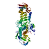
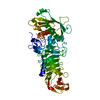
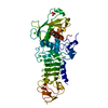
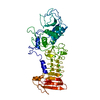
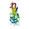
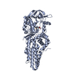
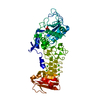
| Title | CRYSTAL STRUCTURE OF A PSYCHROPHILIC ALKALINE PROTEASE FROM PSEUDOMONAS TAC II 18 | ||||||
 Components Components | SERRALYSIN | ||||||
 Keywords Keywords | HYDROLASE / beta jelly roll | ||||||
| Function / homology |  Function and homology information Function and homology informationserralysin / extracellular matrix / metalloendopeptidase activity / calcium ion binding / proteolysis / extracellular space / zinc ion binding Similarity search - Function | ||||||
| Biological species |  Pseudomonas (RNA similarity group I) Pseudomonas (RNA similarity group I) | ||||||
| Method |  X-RAY DIFFRACTION / MOLECULAR REPLACEMENT / Resolution: 1.96 Å X-RAY DIFFRACTION / MOLECULAR REPLACEMENT / Resolution: 1.96 Å | ||||||
 Authors Authors | Aghajari, N. / Haser, R. | ||||||
 Citation Citation | Journal: Proteins / Year: 2003 Title: Crystal structures of a psychrophilic metalloprotease reveal new insights into catalysis by cold-adapted proteases Authors: Aghajari, N. / Van Petegem, F. / Villeret, V. / Chessa, J.P. / Gerday, C. / Haser, R. / Van Beeumen, J. #1: Journal: Embo J. / Year: 1993Title: THREE-DIMENSIONAL STRUCTURE OF THE ALKALINE PROTEASE OF PSEUDOMONAS AERUGINOSA: A TWO-DOMAIN PROTEIN WITH A CALCIUM BINDING PARALLEL BETA ROLL MOTIF Authors: Baumann, U. / Wu, S. / Flaherty, K.M. / McKay, D.B. | ||||||
| History |
| ||||||
| Remark 999 | SEQUENCE AUTHORS CLAIM THAT THE RESIDUE 22 (33 IN SEQUENCE DATABASE) IS ASP (AND NOT GLU) AS JUDGED ...SEQUENCE AUTHORS CLAIM THAT THE RESIDUE 22 (33 IN SEQUENCE DATABASE) IS ASP (AND NOT GLU) AS JUDGED FROM ELECTRON DENSITY MAP |
- Structure visualization
| Structure viewer | Molecule: MolmilJmol/JSmol |
|---|
- Downloads & links
-Download
| PDBx/mmCIF format | 1g9k.cif.gz | 112.3 KB | Display | PDBx/mmCIF format |
|---|---|---|---|---|
| PDB format | pdb1g9k.ent.gz | 83.9 KB | Display | PDB format |
| PDBx/mmJSON format | 1g9k.json.gz | Tree view | PDBx/mmJSON format | |
| Others |  Other downloads Other downloads |
-Validation report
| Summary document | 1g9k_validation.pdf.gz | 437.2 KB | Display | wwPDB validaton report |
|---|---|---|---|---|
| Full document | 1g9k_full_validation.pdf.gz | 438.9 KB | Display | |
| Data in XML | 1g9k_validation.xml.gz | 21 KB | Display | |
| Data in CIF | 1g9k_validation.cif.gz | 31.8 KB | Display | |
| Arichive directory | https://data.pdbj.org/pub/pdb/validation_reports/g9/1g9kftp://data.pdbj.org/pub/pdb/validation_reports/g9/1g9k | HTTPS FTP |
-Related structure data
| Related structure data |  1h71C  1kapS S: Starting model for refinement C: citing same article ( |
|---|---|
| Similar structure data |
-Links
 PDBj
PDBj


- Assembly
| Deposited unit | 
| ||||||||
|---|---|---|---|---|---|---|---|---|---|
| 1 |
| ||||||||
| 2 | 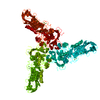
| ||||||||
| Unit cell |
|
-Components
| #1: Protein | Mass: 48727.609 Da / Num. of mol.: 1 / Source method: isolated from a natural source / Source: (natural) Pseudomonas (RNA similarity group I) / Genus: Pseudomonas / Strain: TAC II 18 / References: UniProt: O69771, serralysin | ||||||
|---|---|---|---|---|---|---|---|
| #2: Chemical | ChemComp-CA /   Mass: 40.078 Da / Num. of mol.: 7 / Source method: obtained synthetically / Formula: Ca Mass: 40.078 Da / Num. of mol.: 7 / Source method: obtained synthetically / Formula: Ca#3: Chemical | ChemComp-SO4 / |   Mass: 96.063 Da / Num. of mol.: 1 / Source method: obtained synthetically / Formula: SO4 Mass: 96.063 Da / Num. of mol.: 1 / Source method: obtained synthetically / Formula: SO4#4: Chemical | ChemComp-ZN / |   Mass: 65.409 Da / Num. of mol.: 1 / Source method: obtained synthetically / Formula: Zn Mass: 65.409 Da / Num. of mol.: 1 / Source method: obtained synthetically / Formula: Zn#5: Water | ChemComp-HOH / |  Mass: 18.015 Da / Num. of mol.: 355 / Source method: isolated from a natural source / Formula: H2O Mass: 18.015 Da / Num. of mol.: 355 / Source method: isolated from a natural source / Formula: H2O |
-Experimental details
-Experiment
| Experiment | Method: X-RAY DIFFRACTION / Number of used crystals: 1 |
|---|
- Sample preparation
| Crystal | Density Matthews: 2.59 Å3/Da / Density % sol: 52.58 % | ||||||||||||||||||||||||
|---|---|---|---|---|---|---|---|---|---|---|---|---|---|---|---|---|---|---|---|---|---|---|---|---|---|
| Crystal grow | Temperature: 293 K / Method: vapor diffusion, hanging drop / pH: 7 Details: ammonium sulphate, Hepes, pH 7, VAPOR DIFFUSION, HANGING DROP, temperature 293K | ||||||||||||||||||||||||
| Crystal grow | *PLUS Temperature: 4 ℃ / pH: 7 / Details: Villeret, V., (1997) Protein Sci., 6, 2462. | ||||||||||||||||||||||||
| Components of the solutions | *PLUS
|
-Data collection
| Diffraction | Mean temperature: 289 K |
|---|---|
| Diffraction source | Source: ROTATING ANODE / Type: ENRAF-NONIUS FU581 / Wavelength: 1.5418 Å |
| Detector | Type: MARRESEARCH / Detector: IMAGE PLATE / Date: Oct 20, 2000 / Details: mirrors |
| Radiation | Monochromator: OSMIC MIRRORS / Protocol: SINGLE WAVELENGTH / Monochromatic (M) / Laue (L): M / Scattering type: x-ray |
| Radiation wavelength | Wavelength: 1.5418 Å / Relative weight: 1 |
| Reflection | Resolution: 1.96→93.01 Å / Num. all: 34749 / Num. obs: 33263 / % possible obs: 95.7 % / Observed criterion σ(F): 1 / Observed criterion σ(I): 1 / Redundancy: 4 % / Biso Wilson estimate: 16.1 Å2 / Rmerge(I) obs: 0.058 / Net I/σ(I): 16.1 |
| Reflection shell | Resolution: 1.96→2.01 Å / Redundancy: 2 % / Rmerge(I) obs: 0.268 / Mean I/σ(I) obs: 2.7 / Num. unique all: 2029 / % possible all: 87.6 |
- Processing
| Software |
| |||||||||||||||||||||||||
|---|---|---|---|---|---|---|---|---|---|---|---|---|---|---|---|---|---|---|---|---|---|---|---|---|---|---|
| Refinement | Method to determine structure: MOLECULAR REPLACEMENT Starting model: A lower resolution structure (not yet deposited) solved by the molecular replacement method using pdb entry 1KAP as a search model Resolution: 1.96→46.13 Å Isotropic thermal model: overall anisotropic B-factor correction algorithm from CNS Cross valid method: THROUGHOUT / σ(F): 0 / σ(I): 0 / Stereochemistry target values: Engh & Huber
| |||||||||||||||||||||||||
| Displacement parameters | Biso mean: 18.9 Å2 | |||||||||||||||||||||||||
| Refine analyze |
| |||||||||||||||||||||||||
| Refinement step | Cycle: LAST / Resolution: 1.96→46.13 Å
| |||||||||||||||||||||||||
| Refine LS restraints |
| |||||||||||||||||||||||||
| Refine LS restraints | *PLUS
|
