 Movie
Movie Controller


+ Open data

- Basic information
| Entry | Database: PDB / ID: 1h71 | ||||||
|---|---|---|---|---|---|---|---|
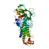
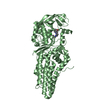
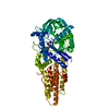
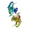
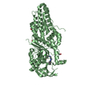
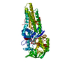
| Title | Psychrophilic Protease from Pseudoalteromonas 'TAC II 18' | ||||||
 Components Components | SERRALYSIN | ||||||
 Keywords Keywords | HYDROLASE / PSYCHROPHILIC / ADAPTATION TO COLD / PROTEASE / DIFFERENT CRYSTAL FORMS | ||||||
| Function / homology |  Function and homology information Function and homology informationserralysin / metalloendopeptidase activity / extracellular matrix / calcium ion binding / proteolysis / : / zinc ion binding Similarity search - Function | ||||||
| Biological species |  PSEUDOMONAS SP. TAC II 18 (bacteria) PSEUDOMONAS SP. TAC II 18 (bacteria) | ||||||
| Method |  X-RAY DIFFRACTION / MOLECULAR REPLACEMENT / Resolution: 2.1 Å X-RAY DIFFRACTION / MOLECULAR REPLACEMENT / Resolution: 2.1 Å | ||||||
 Authors Authors | Villeret, V. / Van Petegem, F. / Aghajari, N. / Chessa, J.-P. / Gerday, C. / Haser, R. / Van Beeumen, J. | ||||||
 Citation Citation | Journal: Proteins: Struct.,Funct., Genet. / Year: 2003 Title: Crystal Structures of a Psychrophilic Metalloprotease Reveal New Insights Into Catalysis by Cold-Adapted Proteases Authors: Aghajari, N. / Van Petegem, F. / Villeret, V. / Chessa, J.-P. / Gerday, C. / Haser, R. / Van Beeumen, J. | ||||||
| History |
|
- Structure visualization
| Structure viewer | Molecule: MolmilJmol/JSmol |
|---|
- Downloads & links
-Download
| PDBx/mmCIF format | 1h71.cif.gz | 108.2 KB | Display | PDBx/mmCIF format |
|---|---|---|---|---|
| PDB format | pdb1h71.ent.gz | 80.1 KB | Display | PDB format |
| PDBx/mmJSON format | 1h71.json.gz | Tree view | PDBx/mmJSON format | |
| Others |  Other downloads Other downloads |
-Validation report
| Arichive directory | https://data.pdbj.org/pub/pdb/validation_reports/h7/1h71ftp://data.pdbj.org/pub/pdb/validation_reports/h7/1h71 | HTTPS FTP |
|---|
-Related structure data
| Related structure data |  1g9kC  1kapS S: Starting model for refinement C: citing same article ( |
|---|---|
| Similar structure data |


-Links
 PDBj
PDBj


- Assembly
| Deposited unit | 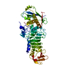
| ||||||||
|---|---|---|---|---|---|---|---|---|---|
| 1 |
| ||||||||
| Unit cell |
|
-Components
| #1: Protein | Mass: 48724.543 Da / Num. of mol.: 1 / Fragment: RESIDUES 18-480 / Source method: isolated from a natural source / Source: (natural) PSEUDOMONAS SP. TAC II 18 (bacteria) / References: UniProt: O69771, serralysin | ||||
|---|---|---|---|---|---|
| #2: Chemical | ChemComp-CA /   Mass: 40.078 Da / Num. of mol.: 8 / Source method: obtained synthetically / Formula: Ca Mass: 40.078 Da / Num. of mol.: 8 / Source method: obtained synthetically / Formula: Ca#3: Chemical | ChemComp-ZN / |   Mass: 65.409 Da / Num. of mol.: 1 / Source method: obtained synthetically / Formula: Zn Mass: 65.409 Da / Num. of mol.: 1 / Source method: obtained synthetically / Formula: Zn#4: Water | ChemComp-HOH / |  Mass: 18.015 Da / Num. of mol.: 284 / Source method: isolated from a natural source / Formula: H2O Mass: 18.015 Da / Num. of mol.: 284 / Source method: isolated from a natural source / Formula: H2O |
-Experimental details
-Experiment
| Experiment | Method: X-RAY DIFFRACTION / Number of used crystals: 1 |
|---|
- Sample preparation
| Crystal | Density Matthews: 2.58 Å3/Da / Density % sol: 53 % | |||||||||||||||||||||||||||||||||||
|---|---|---|---|---|---|---|---|---|---|---|---|---|---|---|---|---|---|---|---|---|---|---|---|---|---|---|---|---|---|---|---|---|---|---|---|---|
| Crystal grow | pH: 7 / Details: pH 7.00 | |||||||||||||||||||||||||||||||||||
| Crystal grow | *PLUS Temperature: 4 or 10 ℃ / Method: vapor diffusion, hanging drop / Details: Villeret, V., (1997) Protein Sci., 6, 2462. | |||||||||||||||||||||||||||||||||||
| Components of the solutions | *PLUS
|
-Data collection
| Diffraction | Mean temperature: 294 K |
|---|---|
| Diffraction source | Source: ROTATING ANODE / Type: ENRAF-NONIUS FR591 / Wavelength: 1.5418 |
| Detector | Type: MAC Science DIP-2030 / Detector: IMAGE PLATE / Details: DOUBLE MIRROR |
| Radiation | Monochromator: NI FILTER / Protocol: SINGLE WAVELENGTH / Monochromatic (M) / Laue (L): M / Scattering type: x-ray |
| Radiation wavelength | Wavelength: 1.5418 Å / Relative weight: 1 |
| Reflection | Resolution: 2.1→15 Å / Num. obs: 29234 / % possible obs: 96.7 % / Redundancy: 2.5 % / Rmerge(I) obs: 0.07 |
| Reflection shell | Resolution: 2.1→2.17 Å / % possible all: 97.7 |
- Processing
| Software |
| ||||||||||||||||||||
|---|---|---|---|---|---|---|---|---|---|---|---|---|---|---|---|---|---|---|---|---|---|
| Refinement | Method to determine structure: MOLECULAR REPLACEMENT Starting model: PDB ENTRY 1KAP Resolution: 2.1→15 Å / Cross valid method: THROUGHOUT
| ||||||||||||||||||||
| Refinement step | Cycle: LAST / Resolution: 2.1→15 Å
| ||||||||||||||||||||
| Refinement | *PLUS Lowest resolution: 15 Å | ||||||||||||||||||||
| Solvent computation | *PLUS | ||||||||||||||||||||
| Displacement parameters | *PLUS | ||||||||||||||||||||
| Refine LS restraints | *PLUS
|
