 Movie
Movie Controller

[English] 日本語
 Yorodumi
- PDB-5xet: Crystal structure of Mycobacterium tuberculosis methionyl-tRNA sy... -
+ Open data

- Basic information
| Entry | Database: PDB / ID: 5xet | ||||||
|---|---|---|---|---|---|---|---|
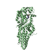
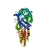
| Title | Crystal structure of Mycobacterium tuberculosis methionyl-tRNA synthetase bound by methionyl-adenylate (Met-AMP) | ||||||
 Components Components | Methionine--tRNA ligase | ||||||
 Keywords Keywords | LIGASE / synthetase / aminoacyl-tRNA synthetase / complex / ATP | ||||||
| Function / homology |  Function and homology information Function and homology informationmethionine-tRNA ligase / methionine-tRNA ligase activity / methionyl-tRNA aminoacylation / ATP binding / metal ion binding / cytoplasm Similarity search - Function | ||||||
| Biological species |  Mycobacterium tuberculosis H37Ra (bacteria) Mycobacterium tuberculosis H37Ra (bacteria) | ||||||
| Method |  X-RAY DIFFRACTION / SYNCHROTRON / MOLECULAR REPLACEMENT / Resolution: 2.38 Å X-RAY DIFFRACTION / SYNCHROTRON / MOLECULAR REPLACEMENT / Resolution: 2.38 Å | ||||||
 Authors Authors | Wang, W. / Qin, B. / Wojdyla, J.A. / Wang, M. / Gao, X. / Cui, S. | ||||||
 Citation Citation | Journal: IUCrJ / Year: 2018 Title: Structural characterization of free-state and product-stateMycobacterium tuberculosismethionyl-tRNA synthetase reveals an induced-fit ligand-recognition mechanism. Authors: Wang, W. / Qin, B. / Wojdyla, J.A. / Wang, M. / Gao, X. / Cui, S. | ||||||
| History |
|
- Structure visualization
| Structure viewer | Molecule: MolmilJmol/JSmol |
|---|
- Downloads & links
-Download
| PDBx/mmCIF format | 5xet.cif.gz | 199.3 KB | Display | PDBx/mmCIF format |
|---|---|---|---|---|
| PDB format | pdb5xet.ent.gz | 157.8 KB | Display | PDB format |
| PDBx/mmJSON format | 5xet.json.gz | Tree view | PDBx/mmJSON format | |
| Others |  Other downloads Other downloads |
-Validation report
| Arichive directory | https://data.pdbj.org/pub/pdb/validation_reports/xe/5xetftp://data.pdbj.org/pub/pdb/validation_reports/xe/5xet | HTTPS FTP |
|---|
-Related structure data
| Related structure data | 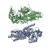 5xgqC 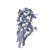 2x1lS S: Starting model for refinement C: citing same article ( |
|---|---|
| Similar structure data |


-Links
 PDBj
PDBj
- Assembly
| Deposited unit | 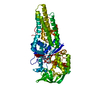
| ||||||||
|---|---|---|---|---|---|---|---|---|---|
| 1 |
| ||||||||
| Unit cell |
|
-Components
| #1: Protein | Mass: 59419.816 Da / Num. of mol.: 1 Source method: isolated from a genetically manipulated source Source: (gene. exp.) Mycobacterium tuberculosis H37Ra (bacteria)Strain: ATCC 25177 / H37Ra / Gene: metG, MRA_1016 Production host: Strain (production host): BL21-Gold(DE3)pLysS AG / References: UniProt: A5U150, methionine-tRNA ligase | ||
|---|---|---|---|
| #2: Chemical | ChemComp-ME8 / [[(  Mass: 478.417 Da / Num. of mol.: 1 Mass: 478.417 Da / Num. of mol.: 1Source method: isolated from a genetically manipulated source Formula: C15H23N6O8PS | ||
| #3: Chemical | ChemComp-EDO /   Mass: 62.068 Da / Num. of mol.: 1 / Source method: isolated from a natural source / Formula: C2H6O2 Mass: 62.068 Da / Num. of mol.: 1 / Source method: isolated from a natural source / Formula: C2H6O2 | ||
| #4: Chemical | ChemComp-MG /   Mass: 24.305 Da / Num. of mol.: 5 / Source method: obtained synthetically / Formula: Mg Mass: 24.305 Da / Num. of mol.: 5 / Source method: obtained synthetically / Formula: Mg#5: Water | ChemComp-HOH / |  Mass: 18.015 Da / Num. of mol.: 180 / Source method: isolated from a natural source / Formula: H2O Mass: 18.015 Da / Num. of mol.: 180 / Source method: isolated from a natural source / Formula: H2O |
-Experimental details
-Experiment
| Experiment | Method: X-RAY DIFFRACTION / Number of used crystals: 1 |
|---|
- Sample preparation
| Crystal | Density Matthews: 2.49 Å3/Da / Density % sol: 50.54 % |
|---|---|
| Crystal grow | Temperature: 293 K / Method: evaporation / pH: 6.9 Details: 0.2M Lithium sulfate monohydrate, 19% PEG 3350, bis tris |
-Data collection
| Diffraction | Mean temperature: 100 K |
|---|---|
| Diffraction source | Source: SYNCHROTRON / Site: SSRF  / Beamline: BL18U1 / Wavelength: 0.97776 Å / Beamline: BL18U1 / Wavelength: 0.97776 Å |
| Detector | Type: DECTRIS PILATUS 6M / Detector: PIXEL / Date: Oct 1, 2016 |
| Radiation | Protocol: SINGLE WAVELENGTH / Monochromatic (M) / Laue (L): M / Scattering type: x-ray |
| Radiation wavelength | Wavelength: 0.97776 Å / Relative weight: 1 |
| Reflection | Resolution: 2.38→37.42 Å / Num. obs: 49191 / % possible obs: 89.8 % / Observed criterion σ(I): -3 / Redundancy: 1.9 % / Biso Wilson estimate: 45.55 Å2 / CC1/2: 0.973 / Rmerge(I) obs: 0.156 / Net I/σ(I): 4.77 |
| Reflection shell | Resolution: 2.38→2.54 Å / Redundancy: 2.13 % / Rmerge(I) obs: 0.585 / Mean I/σ(I) obs: 1.61 / Num. unique obs: 8103 / CC1/2: 0.68 / % possible all: 97.6 |
- Processing
| Software |
| |||||||||||||||||||||||||||||||||||||||||||||||||||||||||||||||
|---|---|---|---|---|---|---|---|---|---|---|---|---|---|---|---|---|---|---|---|---|---|---|---|---|---|---|---|---|---|---|---|---|---|---|---|---|---|---|---|---|---|---|---|---|---|---|---|---|---|---|---|---|---|---|---|---|---|---|---|---|---|---|---|---|
| Refinement | Method to determine structure: MOLECULAR REPLACEMENT Starting model: 2x1l Resolution: 2.38→37.42 Å / SU ML: 0.3 / Cross valid method: THROUGHOUT / σ(F): 1.98 / Phase error: 27.08 Details: THE STRUCTURE FACTOR FILE CONTAINS FRIEDEL PAIRS IN F_PLUS/MINUS AND I_PLUS/MINUS COLUMNS
| |||||||||||||||||||||||||||||||||||||||||||||||||||||||||||||||
| Solvent computation | Shrinkage radii: 0.9 Å / VDW probe radii: 1.11 Å | |||||||||||||||||||||||||||||||||||||||||||||||||||||||||||||||
| Refinement step | Cycle: LAST / Resolution: 2.38→37.42 Å
| |||||||||||||||||||||||||||||||||||||||||||||||||||||||||||||||
| Refine LS restraints |
| |||||||||||||||||||||||||||||||||||||||||||||||||||||||||||||||
| LS refinement shell |
|
