| Entry | Database: PDB / ID: 6ax8
|
|---|
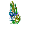
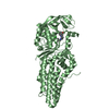
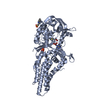
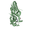
| Title | Mycobacterium tuberculosis methionyl-tRNA synthetase in complex with methionyl-adenylate |
|---|
 Components Components | Methionine-tRNA ligase |
|---|
 Keywords Keywords | LIGASE / synthetase / Ligase-aminoacyl adenylate complex |
|---|
| Function / homology |  Function and homology information Function and homology information
methionine-tRNA ligase / methionine-tRNA ligase activity / methionyl-tRNA aminoacylation / ATP binding / plasma membrane / cytoplasmSimilarity search - Function Methionyl-trna Synthetase; domain 2 / Methionyl-trna Synthetase; domain 2 - #10 / Methionine-tRNA synthetase, type 2 / Anticodon binding domain of methionyl tRNA ligase / Methionyl-tRNA synthetase / Methioninyl-tRNA synthetase core domain / Methionyl-tRNA synthetase, anticodon-binding domain / Isoleucyl-tRNA Synthetase; Domain 1 / Isoleucyl-tRNA Synthetase; Domain 1 / Methionyl/Leucyl tRNA synthetase ...Methionyl-trna Synthetase; domain 2 / Methionyl-trna Synthetase; domain 2 - #10 / Methionine-tRNA synthetase, type 2 / Anticodon binding domain of methionyl tRNA ligase / Methionyl-tRNA synthetase / Methioninyl-tRNA synthetase core domain / Methionyl-tRNA synthetase, anticodon-binding domain / Isoleucyl-tRNA Synthetase; Domain 1 / Isoleucyl-tRNA Synthetase; Domain 1 / Methionyl/Leucyl tRNA synthetase / tRNA synthetases class I (M) / Aminoacyl-tRNA synthetase, class Ia, anticodon-binding / Aminoacyl-tRNA synthetase, class I, conserved site / Aminoacyl-transfer RNA synthetases class-I signature. / HUPs / Rossmann-like alpha/beta/alpha sandwich fold / Beta Complex / Rossmann fold / Orthogonal Bundle / 3-Layer(aba) Sandwich / Mainly Beta / Mainly Alpha / Alpha BetaSimilarity search - Domain/homology |
|---|
| Biological species |   Mycobacterium tuberculosis (bacteria) Mycobacterium tuberculosis (bacteria) |
|---|
| Method |  X-RAY DIFFRACTION / SYNCHROTRON / MOLECULAR REPLACEMENT / Resolution: 2.6 Å X-RAY DIFFRACTION / SYNCHROTRON / MOLECULAR REPLACEMENT / Resolution: 2.6 Å |
|---|
 Authors Authors | Barros-Alvarez, X. / Hol, W.G.J. |
|---|
| Funding support |  United States, 1items United States, 1items | Organization | Grant number | Country |
|---|
| National Institutes of Health/National Institute Of Allergy and Infectious Diseases (NIH/NIAID) | RO1 AI084004 and RO1 AI097177 | United States |
|
|---|
 Citation Citation | Journal: Acta Crystallogr F Struct Biol Commun / Year: 2018
Title: The crystal structure of the drug target Mycobacterium tuberculosis methionyl-tRNA synthetase in complex with a catalytic intermediate.
Authors: Barros-Alvarez, X. / Turley, S. / Ranade, R.M. / Gillespie, J.R. / Duster, N.A. / Verlinde, C.L.M.J. / Fan, E. / Buckner, F.S. / Hol, W.G.J. |
|---|
| History | | Deposition | Sep 6, 2017 | Deposition site: RCSB / Processing site: RCSB |
|---|
| Revision 1.0 | Apr 11, 2018 | Provider: repository / Type: Initial release |
|---|
| Revision 1.1 | Oct 31, 2018 | Group: Data collection / Database references / Category: citation
Item: _citation.journal_abbrev / _citation.pdbx_database_id_PubMed / _citation.title |
|---|
| Revision 1.2 | Feb 20, 2019 | Group: Author supporting evidence / Data collection / Category: pdbx_audit_support / Item: _pdbx_audit_support.funding_organization |
|---|
| Revision 1.3 | Dec 11, 2019 | Group: Author supporting evidence / Category: pdbx_audit_support / Item: _pdbx_audit_support.funding_organization |
|---|
| Revision 1.4 | Mar 13, 2024 | Group: Data collection / Database references / Category: chem_comp_atom / chem_comp_bond / database_2
Item: _database_2.pdbx_DOI / _database_2.pdbx_database_accession |
|---|
|
|---|
 Movie
Movie Controller

 Yorodumi
 Open data
 Basic information
 Structure visualization
 Downloads & links
 Other downloads

 PDBj
PDBj
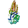
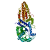
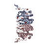
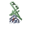
 Assembly
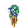
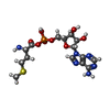
 Mass: 478.417 Da / Num. of mol.: 1 / Source method: obtained synthetically / Formula: C15H23N6O8PS / Feature type: SUBJECT OF INVESTIGATION
 Mass: 18.015 Da / Num. of mol.: 38 / Source method: isolated from a natural source / Formula: H2O
 Sample preparation
 Processing