 Movie
Movie Controller

[English] 日本語
 Yorodumi
- PDB-2x1l: Crystal structure of Mycobacterium smegmatis methionyl-tRNA synth... -
+ Open data

- Basic information
| Entry | Database: PDB / ID: 2x1l | ||||||
|---|---|---|---|---|---|---|---|
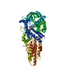
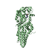
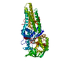
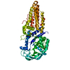
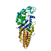
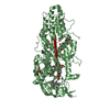
| Title | Crystal structure of Mycobacterium smegmatis methionyl-tRNA synthetase in complex with methionine and adenosine | ||||||
 Components Components | METHIONYL-TRNA SYNTHETASE | ||||||
 Keywords Keywords | LIGASE / NUCLEOTIDE-BINDING / PROTEIN BIOSYNTHESIS / AMINOACYL-TRNA SYNTHETASE | ||||||
| Function / homology |  Function and homology information Function and homology informationmethionine-tRNA ligase / methionine-tRNA ligase activity / methionyl-tRNA aminoacylation / ATP binding / cytoplasm Similarity search - Function | ||||||
| Biological species |  MYCOBACTERIUM SMEGMATIS (bacteria) MYCOBACTERIUM SMEGMATIS (bacteria) | ||||||
| Method |  X-RAY DIFFRACTION / SYNCHROTRON / MOLECULAR REPLACEMENT / Resolution: 2.3 Å X-RAY DIFFRACTION / SYNCHROTRON / MOLECULAR REPLACEMENT / Resolution: 2.3 Å | ||||||
 Authors Authors | Ingvarsson, H. / Jones, T.A. / Unge, T. | ||||||
 Citation Citation | Journal: FEBS J. / Year: 2010 Title: Flexibility and Communication within the Structure of the Mycobacterium Smegmatis Methionyl-tRNA Synthetase. Authors: Ingvarsson, H. / Unge, T. #1: Journal: Acta Crystallogr.,Sect.F / Year: 2009 Title: Crystallization of Mycobacterium Smegmatis Methionyl-tRNA Synthetase in the Presence of Methionine and Adenosine. Authors: Ingvarsson, H. / Jones, T.A. / Unge, T. | ||||||
| History |
|
- Structure visualization
| Structure viewer | Molecule: MolmilJmol/JSmol |
|---|
- Downloads & links
-Download
| PDBx/mmCIF format | 2x1l.cif.gz | 304.7 KB | Display | PDBx/mmCIF format |
|---|---|---|---|---|
| PDB format | pdb2x1l.ent.gz | 245.1 KB | Display | PDB format |
| PDBx/mmJSON format | 2x1l.json.gz | Tree view | PDBx/mmJSON format | |
| Others |  Other downloads Other downloads |
-Validation report
| Arichive directory | https://data.pdbj.org/pub/pdb/validation_reports/x1/2x1lftp://data.pdbj.org/pub/pdb/validation_reports/x1/2x1l | HTTPS FTP |
|---|
-Related structure data
| Related structure data |  2x1mC 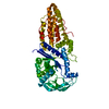 1a8hS C: citing same article ( S: Starting model for refinement |
|---|---|
| Similar structure data |


-Links
 PDBj
PDBj

- Assembly
| Deposited unit | 
| ||||||||
|---|---|---|---|---|---|---|---|---|---|
| 1 | 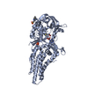
| ||||||||
| 2 | 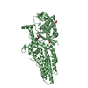
| ||||||||
| 3 | 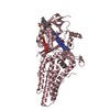
| ||||||||
| Unit cell |
|
-Components
-Protein , 1 types, 3 molecules ABC
| #1: Protein | Mass: 59175.215 Da / Num. of mol.: 3 / Fragment: RESIDUES 2-515 Source method: isolated from a genetically manipulated source Source: (gene. exp.) MYCOBACTERIUM SMEGMATIS (bacteria) / Strain: MC2 155 / Production host: |
|---|
-Non-polymers , 5 types, 496 molecules 


| #2: Chemical |  Type: L-peptide linking / Mass: 149.211 Da / Num. of mol.: 3 / Source method: obtained synthetically / Formula: C5H11NO2S Type: L-peptide linking / Mass: 149.211 Da / Num. of mol.: 3 / Source method: obtained synthetically / Formula: C5H11NO2S#3: Chemical |  Mass: 267.241 Da / Num. of mol.: 3 / Source method: obtained synthetically / Formula: C10H13N5O4 Mass: 267.241 Da / Num. of mol.: 3 / Source method: obtained synthetically / Formula: C10H13N5O4#4: Chemical |  Mass: 221.317 Da / Num. of mol.: 3 / Source method: obtained synthetically / Formula: C9H19NO3S / Comment: pH buffer*YM Mass: 221.317 Da / Num. of mol.: 3 / Source method: obtained synthetically / Formula: C9H19NO3S / Comment: pH buffer*YM#5: Chemical |  Mass: 96.987 Da / Num. of mol.: 3 / Source method: obtained synthetically / Formula: H2O4P Mass: 96.987 Da / Num. of mol.: 3 / Source method: obtained synthetically / Formula: H2O4P#6: Water | ChemComp-HOH / | Mass: 18.015 Da / Num. of mol.: 484 / Source method: isolated from a natural source / Formula: H2O |
|---|
-Experimental details
-Experiment
| Experiment | Method: X-RAY DIFFRACTION / Number of used crystals: 1 |
|---|
- Sample preparation
| Crystal | Density Matthews: 3.2 Å3/Da / Density % sol: 61 % / Description: NONE |
|---|---|
| Crystal grow | Temperature: 293 K / Method: vapor diffusion, hanging drop / pH: 7 Details: 1.2 M NAH2PO4, 800 MM K2HPO4, 200 MM LI2SO4, 100 MM CAPS, PH 7.0, VAPOR DIFFUSION, HANGING DROPS, TEMPERATURE 293 K |
-Data collection
| Diffraction | Mean temperature: 100 K |
|---|---|
| Diffraction source | Source: SYNCHROTRON / Site: MAX II  / Beamline: I911-2 / Wavelength: 1.038 / Beamline: I911-2 / Wavelength: 1.038 |
| Detector | Type: MARRESEARCH / Detector: CCD / Date: Oct 10, 2008 / Details: MULTILAYER MIRROR |
| Radiation | Monochromator: BENT SI (111) CRYSTAL, HORIZONTALLY FOCUSING / Protocol: SINGLE WAVELENGTH / Monochromatic (M) / Laue (L): M / Scattering type: x-ray |
| Radiation wavelength | Wavelength: 1.038 Å / Relative weight: 1 |
| Reflection | Resolution: 2.3→30 Å / Num. obs: 93306 / % possible obs: 97.8 % / Observed criterion σ(I): 2 / Redundancy: 4.1 % / Biso Wilson estimate: 39.6 Å2 / Rmerge(I) obs: 0.09 / Net I/σ(I): 11.8 |
| Reflection shell | Resolution: 2.3→2.42 Å / Redundancy: 3.8 % / Rmerge(I) obs: 0.38 / Mean I/σ(I) obs: 3.2 / % possible all: 96.1 |
- Processing
| Software |
| ||||||||||||||||||||||||||||||||||||||||||||||||||||||||||||||||||||||||||||||||||||||||||||||||||||||||||||||||||||||||||||||||||||||||||||||||||||||||||||||||||||||||||||||||||||||
|---|---|---|---|---|---|---|---|---|---|---|---|---|---|---|---|---|---|---|---|---|---|---|---|---|---|---|---|---|---|---|---|---|---|---|---|---|---|---|---|---|---|---|---|---|---|---|---|---|---|---|---|---|---|---|---|---|---|---|---|---|---|---|---|---|---|---|---|---|---|---|---|---|---|---|---|---|---|---|---|---|---|---|---|---|---|---|---|---|---|---|---|---|---|---|---|---|---|---|---|---|---|---|---|---|---|---|---|---|---|---|---|---|---|---|---|---|---|---|---|---|---|---|---|---|---|---|---|---|---|---|---|---|---|---|---|---|---|---|---|---|---|---|---|---|---|---|---|---|---|---|---|---|---|---|---|---|---|---|---|---|---|---|---|---|---|---|---|---|---|---|---|---|---|---|---|---|---|---|---|---|---|---|---|
| Refinement | Method to determine structure: MOLECULAR REPLACEMENT Starting model: PDB ENTRY 1A8H Resolution: 2.3→30 Å / Cor.coef. Fo:Fc: 0.921 / Cor.coef. Fo:Fc free: 0.899 / SU B: 6.18 / SU ML: 0.15 / Cross valid method: THROUGHOUT / ESU R: 0.29 / ESU R Free: 0.22 / Stereochemistry target values: MAXIMUM LIKELIHOOD Details: HYDROGENS HAVE BEEN ADDED IN THE RIDING POSITIONS. RESIDUES 124 TO 160 AND 123 TO 158 ARE DISORDERED IN MOLECULE B AND C, RESPECTIVELY
| ||||||||||||||||||||||||||||||||||||||||||||||||||||||||||||||||||||||||||||||||||||||||||||||||||||||||||||||||||||||||||||||||||||||||||||||||||||||||||||||||||||||||||||||||||||||
| Solvent computation | Ion probe radii: 0.8 Å / Shrinkage radii: 0.8 Å / VDW probe radii: 1.4 Å / Solvent model: MASK | ||||||||||||||||||||||||||||||||||||||||||||||||||||||||||||||||||||||||||||||||||||||||||||||||||||||||||||||||||||||||||||||||||||||||||||||||||||||||||||||||||||||||||||||||||||||
| Displacement parameters | Biso mean: 23.9 Å2
| ||||||||||||||||||||||||||||||||||||||||||||||||||||||||||||||||||||||||||||||||||||||||||||||||||||||||||||||||||||||||||||||||||||||||||||||||||||||||||||||||||||||||||||||||||||||
| Refinement step | Cycle: LAST / Resolution: 2.3→30 Å
| ||||||||||||||||||||||||||||||||||||||||||||||||||||||||||||||||||||||||||||||||||||||||||||||||||||||||||||||||||||||||||||||||||||||||||||||||||||||||||||||||||||||||||||||||||||||
| Refine LS restraints |
|