 Movie
Movie Controller

[English] 日本語
 Yorodumi
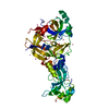
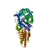
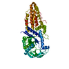
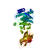
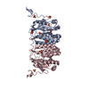
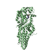
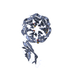
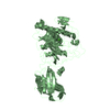
- PDB-3tkl: Crystal structure of the GTP-bound Rab1a in complex with the coil... -
+ Open data

- Basic information
| Entry | Database: PDB / ID: 3tkl | ||||||
|---|---|---|---|---|---|---|---|
| Title | Crystal structure of the GTP-bound Rab1a in complex with the coiled-coil domain of LidA from Legionella pneumophila | ||||||
 Components Components |
| ||||||
 Keywords Keywords | PROTEIN TRANSPORT/PROTEIN BINDING / Vesicle trafficking / PROTEIN TRANSPORT-PROTEIN BINDING complex | ||||||
| Function / homology |  Function and homology information Function and homology informationpositive regulation of glycoprotein metabolic process / growth hormone secretion / COPII-coated vesicle cargo loading / RAB geranylgeranylation / melanosome transport / RAB GEFs exchange GTP for GDP on RABs / Golgi Cisternae Pericentriolar Stack Reorganization / vesicle transport along microtubule / COPII-mediated vesicle transport / COPI-dependent Golgi-to-ER retrograde traffic ...positive regulation of glycoprotein metabolic process / growth hormone secretion / COPII-coated vesicle cargo loading / RAB geranylgeranylation / melanosome transport / RAB GEFs exchange GTP for GDP on RABs / Golgi Cisternae Pericentriolar Stack Reorganization / vesicle transport along microtubule / COPII-mediated vesicle transport / COPI-dependent Golgi-to-ER retrograde traffic / virion assembly / transport vesicle membrane / Golgi organization / endoplasmic reticulum to Golgi vesicle-mediated transport / autophagosome assembly / COPI-mediated anterograde transport / vesicle-mediated transport / endomembrane system / substrate adhesion-dependent cell spreading / positive regulation of interleukin-8 production / small monomeric GTPase / intracellular protein transport / autophagy / endocytosis / melanosome / cell migration / G protein activity / early endosome / defense response to bacterium / cadherin binding / Golgi membrane / GTPase activity / GTP binding / Golgi apparatus / endoplasmic reticulum / extracellular exosome / metal ion binding / cytosol Similarity search - Function | ||||||
| Biological species |  Homo sapiens (human) Homo sapiens (human)  Legionella pneumophila (bacteria) Legionella pneumophila (bacteria) | ||||||
| Method |  X-RAY DIFFRACTION / SYNCHROTRON / MOLECULAR REPLACEMENT / Resolution: 2.183 Å X-RAY DIFFRACTION / SYNCHROTRON / MOLECULAR REPLACEMENT / Resolution: 2.183 Å | ||||||
 Authors Authors | Cheng, W. / Yin, K. / Lu, D. / Li, B. / Zhu, D. / Chen, Y. / Zhang, H. / Xu, S. / Chai, J. / Gu, L. | ||||||
 Citation Citation | Journal: Plos Pathog. / Year: 2012 Title: Structural insights into a unique Legionella pneumophila effector LidA recognizing both GDP and GTP bound Rab1 in their active state Authors: Cheng, W. / Yin, K. / Lu, D. / Li, B. / Zhu, D. / Chen, Y. / Zhang, H. / Xu, S. / Chai, J. / Gu, L. | ||||||
| History |
|
- Structure visualization
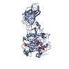
| Structure viewer | Molecule: MolmilJmol/JSmol |
|---|
- Downloads & links
-Download
| PDBx/mmCIF format | 3tkl.cif.gz | 92.3 KB | Display | PDBx/mmCIF format |
|---|---|---|---|---|
| PDB format | pdb3tkl.ent.gz | 66.5 KB | Display | PDB format |
| PDBx/mmJSON format | 3tkl.json.gz | Tree view | PDBx/mmJSON format | |
| Others |  Other downloads Other downloads |
-Validation report
| Arichive directory | https://data.pdbj.org/pub/pdb/validation_reports/tk/3tklftp://data.pdbj.org/pub/pdb/validation_reports/tk/3tkl | HTTPS FTP |
|---|
-Related structure data
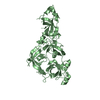
| Related structure data | 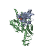 3sfvC 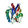 2folS C: citing same article ( S: Starting model for refinement |
|---|---|
| Similar structure data |
-Links
 PDBj
PDBj


- Assembly
| Deposited unit | 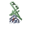
| ||||||||
|---|---|---|---|---|---|---|---|---|---|
| 1 |
| ||||||||
| Unit cell |
|
-Components
| #1: Protein | Mass: 21766.635 Da / Num. of mol.: 1 / Fragment: UNP residues 1-191 Source method: isolated from a genetically manipulated source Source: (gene. exp.) Homo sapiens (human) / Gene: RAB1 / Plasmid: pGEX-6P-1 / Production host: |
|---|---|
| #2: Protein | Mass: 30634.645 Da / Num. of mol.: 1 / Fragment: Coiled-coil domain Source method: isolated from a genetically manipulated source Source: (gene. exp.) Legionella pneumophila (bacteria) / Strain: Corby / Gene: lidA / Plasmid: pGEX-6P-1 / Production host: |
| #3: Chemical | ChemComp-MG /   Mass: 24.305 Da / Num. of mol.: 1 / Source method: obtained synthetically / Formula: Mg Mass: 24.305 Da / Num. of mol.: 1 / Source method: obtained synthetically / Formula: Mg |
| #4: Chemical | ChemComp-GTP /   Mass: 523.180 Da / Num. of mol.: 1 / Source method: obtained synthetically / Formula: C10H16N5O14P3 / Comment: GTP, energy-carrying molecule*YM Mass: 523.180 Da / Num. of mol.: 1 / Source method: obtained synthetically / Formula: C10H16N5O14P3 / Comment: GTP, energy-carrying molecule*YM |
| #5: Water | ChemComp-HOH /  Mass: 18.015 Da / Num. of mol.: 111 / Source method: isolated from a natural source / Formula: H2O Mass: 18.015 Da / Num. of mol.: 111 / Source method: isolated from a natural source / Formula: H2O |
-Experimental details
-Experiment
| Experiment | Method: X-RAY DIFFRACTION / Number of used crystals: 1 |
|---|
- Sample preparation
| Crystal | Density Matthews: 1.86 Å3/Da / Density % sol: 33.89 % |
|---|---|
| Crystal grow | Temperature: 293 K / Method: vapor diffusion, hanging drop / pH: 7.5 Details: 0.1M HEPES, 25% PEG3350, 0.7% butanol, pH 7.5, VAPOR DIFFUSION, HANGING DROP, temperature 293K |
-Data collection
| Diffraction | Mean temperature: 100 K |
|---|---|
| Diffraction source | Source: SYNCHROTRON / Site: SSRF  / Beamline: BL17U / Wavelength: 1 Å / Beamline: BL17U / Wavelength: 1 Å |
| Detector | Type: RAYONIX MX-225 / Detector: CCD / Date: Jun 14, 2010 |
| Radiation | Monochromator: Si(111) / Protocol: SINGLE WAVELENGTH / Monochromatic (M) / Laue (L): M / Scattering type: x-ray |
| Radiation wavelength | Wavelength: 1 Å / Relative weight: 1 |
| Reflection | Resolution: 2.183→50 Å / Num. all: 20017 / Num. obs: 20017 / % possible obs: 99.6 % / Observed criterion σ(F): 0 / Observed criterion σ(I): 0 / Redundancy: 4.2 % / Biso Wilson estimate: 25.74 Å2 / Rmerge(I) obs: 0.091 / Rsym value: 0.091 / Net I/σ(I): 23.88 |
| Reflection shell | Resolution: 2.2→2.24 Å / Redundancy: 3.9 % / Rmerge(I) obs: 0.295 / Mean I/σ(I) obs: 4.65 / Num. unique all: 991 / Rsym value: 0.295 / % possible all: 98.7 |
- Processing
| Software |
| ||||||||||||||||||||||||||||||||||||||||||||||||||||||||
|---|---|---|---|---|---|---|---|---|---|---|---|---|---|---|---|---|---|---|---|---|---|---|---|---|---|---|---|---|---|---|---|---|---|---|---|---|---|---|---|---|---|---|---|---|---|---|---|---|---|---|---|---|---|---|---|---|---|
| Refinement | Method to determine structure: MOLECULAR REPLACEMENT Starting model: PDB ENTRY 2FOL Resolution: 2.183→31.785 Å / Occupancy max: 1 / Occupancy min: 1 / SU ML: 0.25 / σ(F): 1.35 / σ(I): 0 / Phase error: 25.08 / Stereochemistry target values: ML
| ||||||||||||||||||||||||||||||||||||||||||||||||||||||||
| Solvent computation | Shrinkage radii: 0.9 Å / VDW probe radii: 1.11 Å / Solvent model: FLAT BULK SOLVENT MODEL / Bsol: 37.449 Å2 / ksol: 0.372 e/Å3 | ||||||||||||||||||||||||||||||||||||||||||||||||||||||||
| Displacement parameters | Biso max: 89.92 Å2 / Biso mean: 32.0164 Å2 / Biso min: 14.28 Å2
| ||||||||||||||||||||||||||||||||||||||||||||||||||||||||
| Refinement step | Cycle: LAST / Resolution: 2.183→31.785 Å
| ||||||||||||||||||||||||||||||||||||||||||||||||||||||||
| Refine LS restraints |
| ||||||||||||||||||||||||||||||||||||||||||||||||||||||||
| LS refinement shell | Refine-ID: X-RAY DIFFRACTION / Total num. of bins used: 7
|
