 Movie
Movie Controller

[English] 日本語
 Yorodumi
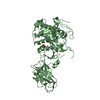
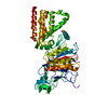
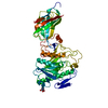
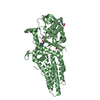
- PDB-1hpl: HORSE PANCREATIC LIPASE. THE CRYSTAL STRUCTURE AT 2.3 ANGSTROMS R... -
+ Open data

- Basic information
| Entry | Database: PDB / ID: 1hpl | ||||||
|---|---|---|---|---|---|---|---|
| Title | HORSE PANCREATIC LIPASE. THE CRYSTAL STRUCTURE AT 2.3 ANGSTROMS RESOLUTION | ||||||
 Components Components | LIPASE | ||||||
 Keywords Keywords | HYDROLASE(CARBOXYLIC ESTERASE) | ||||||
| Function / homology |  Function and homology information Function and homology informationall-trans-retinyl-palmitate hydrolase, all-trans-retinol forming activity / lipoprotein lipase activity / glycerophospholipid phospholipase A1 activity / triglyceride catabolic process / triacylglycerol lipase / high-density lipoprotein particle remodeling / triacylglycerol lipase activity / cholesterol homeostasis / fatty acid biosynthetic process / : / metal ion binding Similarity search - Function | ||||||
| Biological species |  | ||||||
| Method |  X-RAY DIFFRACTION / Resolution: 2.3 Å X-RAY DIFFRACTION / Resolution: 2.3 Å | ||||||
 Authors Authors | Bourne, Y. / Cambillau, C. | ||||||
 Citation Citation | Journal: J.Mol.Biol. / Year: 1994 Title: Horse pancreatic lipase. The crystal structure refined at 2.3 A resolution. Authors: Bourne, Y. / Martinez, C. / Kerfelec, B. / Lombardo, D. / Chapus, C. / Cambillau, C. #1: Journal: J.Mol.Biol. / Year: 1989Title: Crystallization and Preliminary X-Ray Study of Horse Pancreatic Lipase Authors: Lombardo, D. / Chapus, C. / Bourne, Y. / Cambillau, C. | ||||||
| History |
|
- Structure visualization
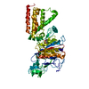
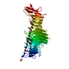
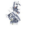
| Structure viewer | Molecule: MolmilJmol/JSmol |
|---|
- Downloads & links
-Download
| PDBx/mmCIF format | 1hpl.cif.gz | 199.4 KB | Display | PDBx/mmCIF format |
|---|---|---|---|---|
| PDB format | pdb1hpl.ent.gz | 157.6 KB | Display | PDB format |
| PDBx/mmJSON format | 1hpl.json.gz | Tree view | PDBx/mmJSON format | |
| Others |  Other downloads Other downloads |
-Validation report
| Arichive directory | https://data.pdbj.org/pub/pdb/validation_reports/hp/1hplftp://data.pdbj.org/pub/pdb/validation_reports/hp/1hpl | HTTPS FTP |
|---|
-Related structure data
| Similar structure data |
|---|

-Links
 PDBj
PDBj- Assembly
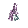
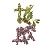
| Deposited unit | 
| ||||||||
|---|---|---|---|---|---|---|---|---|---|
| 1 | 
| ||||||||
| 2 | 
| ||||||||
| Unit cell |
| ||||||||
| Atom site foot note | 1: CIS PROLINE - PRO A 16 / 2: CIS PROLINE - PRO A 211 / 3: CIS PROLINE - PRO A 298 4: SER A 333 - ASN A 334 OMEGA =326.11 PEPTIDE BOND DEVIATES SIGNIFICANTLY FROM TRANS CONFORMATION 5: CIS PROLINE - PRO B 16 / 6: CIS PROLINE - PRO B 211 / 7: CIS PROLINE - PRO B 298 8: SER B 333 - ASN B 334 OMEGA =298.62 PEPTIDE BOND DEVIATES SIGNIFICANTLY FROM TRANS CONFORMATION | ||||||||
| Noncrystallographic symmetry (NCS) | NCS oper: (Code: given Matrix: (-0.9999, -0.0011, 0.0029), Vector: |
-Components
| #1: Protein | Mass: 49759.441 Da / Num. of mol.: 2 Source method: isolated from a genetically manipulated source Source: (gene. exp.) #2: Chemical |   Mass: 40.078 Da / Num. of mol.: 2 / Source method: obtained synthetically / Formula: Ca Mass: 40.078 Da / Num. of mol.: 2 / Source method: obtained synthetically / Formula: Ca#3: Water | ChemComp-HOH / |  Mass: 18.015 Da / Num. of mol.: 705 / Source method: isolated from a natural source / Formula: H2O Mass: 18.015 Da / Num. of mol.: 705 / Source method: isolated from a natural source / Formula: H2OHas protein modification | Y | |
|---|
-Experimental details
-Experiment
| Experiment | Method: X-RAY DIFFRACTION |
|---|
- Sample preparation
| Crystal | Density Matthews: 2.83 Å3/Da / Density % sol: 56.54 % | ||||||||||||||||||||||||||||||||||||
|---|---|---|---|---|---|---|---|---|---|---|---|---|---|---|---|---|---|---|---|---|---|---|---|---|---|---|---|---|---|---|---|---|---|---|---|---|---|
| Crystal grow | *PLUS Temperature: 20 ℃ / pH: 5.6 / Method: vapor diffusion, hanging drop / Details: referred to J.Mol.Biol. 205.259-261 | ||||||||||||||||||||||||||||||||||||
| Components of the solutions | *PLUS
|
-Data collection
| Radiation | Scattering type: x-ray |
|---|---|
| Radiation wavelength | Relative weight: 1 |
| Reflection | *PLUS Highest resolution: 2.3 Å / Num. obs: 45644 / % possible obs: 86.3 % / Num. measured all: 145844 / Rmerge(I) obs: 0.063 |
| Reflection shell | *PLUS Highest resolution: 2.3 Å / Lowest resolution: 2.4 Å / % possible obs: 48 % |
- Processing
| Software |
| ||||||||||||||||||||||||||||||||||||||||||||||||||||||||||||
|---|---|---|---|---|---|---|---|---|---|---|---|---|---|---|---|---|---|---|---|---|---|---|---|---|---|---|---|---|---|---|---|---|---|---|---|---|---|---|---|---|---|---|---|---|---|---|---|---|---|---|---|---|---|---|---|---|---|---|---|---|---|
| Refinement | Resolution: 2.3→6 Å / Rfactor Rwork: 0.159 / Rfactor obs: 0.159 / σ(F): 1 | ||||||||||||||||||||||||||||||||||||||||||||||||||||||||||||
| Refinement step | Cycle: LAST / Resolution: 2.3→6 Å
| ||||||||||||||||||||||||||||||||||||||||||||||||||||||||||||
| Refine LS restraints |
| ||||||||||||||||||||||||||||||||||||||||||||||||||||||||||||
| Software | *PLUS Name: X-PLOR / Classification: refinement | ||||||||||||||||||||||||||||||||||||||||||||||||||||||||||||
| Refinement | *PLUS Rfactor obs: 0.159 / Rfactor Rwork: 0.159 | ||||||||||||||||||||||||||||||||||||||||||||||||||||||||||||
| Solvent computation | *PLUS | ||||||||||||||||||||||||||||||||||||||||||||||||||||||||||||
| Displacement parameters | *PLUS | ||||||||||||||||||||||||||||||||||||||||||||||||||||||||||||
| Refine LS restraints | *PLUS Type: x_angle_d / Dev ideal: 3.1 |
