 Movie
Movie Controller

[English] 日本語
 Yorodumi
- PDB-1kix: Dimeric Structure of the O. nova Telomere End Binding Protein Alp... -
+ Open data

- Basic information
| Entry | Database: PDB / ID: 1kix | ||||||
|---|---|---|---|---|---|---|---|
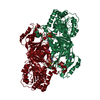
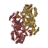
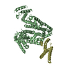
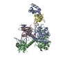
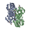
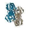
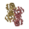
| Title | Dimeric Structure of the O. nova Telomere End Binding Protein Alpha Subunit with Bound ssDNA | ||||||
 Components Components |
| ||||||
 Keywords Keywords | DNA BINDING PROTEIN/DNA / TELOMERE BINDING PROTEIN / DNA-PROTEIN INTERACTIONS / SINGLE STRANDED DNA / ssDNA / DNA BINDING PROTEIN-DNA COMPLEX | ||||||
| Function / homology |  Function and homology information Function and homology informationtelomere cap complex / telomerase inhibitor activity / regulation of telomere maintenance via telomerase / single-stranded telomeric DNA binding / G-rich strand telomeric DNA binding / nuclear telomere cap complex / telomere capping / protein-containing complex Similarity search - Function | ||||||
| Biological species |  Sterkiella nova (eukaryote) Sterkiella nova (eukaryote) | ||||||
| Method |  X-RAY DIFFRACTION / SYNCHROTRON / MOLECULAR REPLACEMENT / Resolution: 2.7 Å X-RAY DIFFRACTION / SYNCHROTRON / MOLECULAR REPLACEMENT / Resolution: 2.7 Å | ||||||
 Authors Authors | Peersen, O.B. / Ruggles, J.A. / Schultz, S.C. | ||||||
 Citation Citation | Journal: Nat.Struct.Biol. / Year: 2002 Title: Dimeric structure of the Oxytricha nova telomere end-binding protein alpha-subunit bound to ssDNA. Authors: Peersen, O.B. / Ruggles, J.A. / Schultz, S.C. #1: Journal: Cell(Cambridge,Mass.) / Year: 1998Title: Crystal structure of the Oxytricha nova telomere end binding protein complexed with single strand DNA Authors: Horvath, M.P. / Schweiker, V.L. / Bevilacqua, J.M. / Ruggles, J.A. / Schultz, S.C. #2: Journal: J.Mol.Biol. / Year: 2001Title: Crystal Structure of the N-terminal Domain of Oxytricha nova Telomere End-binding Protein alpha Subunit both Uncomplexed and Complexed with Telomeric ssDNA Authors: Classen, S. / Ruggles, J.A. / Schultz, S.C. | ||||||
| History |
|
- Structure visualization
| Structure viewer | Molecule: MolmilJmol/JSmol |
|---|
- Downloads & links
-Download
| PDBx/mmCIF format | 1kix.cif.gz | 110.8 KB | Display | PDBx/mmCIF format |
|---|---|---|---|---|
| PDB format | pdb1kix.ent.gz | 84.6 KB | Display | PDB format |
| PDBx/mmJSON format | 1kix.json.gz | Tree view | PDBx/mmJSON format | |
| Others |  Other downloads Other downloads |
-Validation report
| Arichive directory | https://data.pdbj.org/pub/pdb/validation_reports/ki/1kixftp://data.pdbj.org/pub/pdb/validation_reports/ki/1kix | HTTPS FTP |
|---|
-Related structure data
| Related structure data |  1otcS S: Starting model for refinement |
|---|---|
| Similar structure data |


-Links
 PDBj
PDBj- Assembly
| Deposited unit | 
| ||||||||
|---|---|---|---|---|---|---|---|---|---|
| 1 | 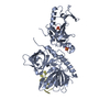
| ||||||||
| Unit cell |
|
-Components
| #1: DNA chain | Mass: 2488.637 Da / Num. of mol.: 1 / Source method: obtained synthetically Details: 3' Terminal Single Strand DNA Sequence of Macronuclear Telomeres | ||
|---|---|---|---|
| #2: Protein | Mass: 56168.301 Da / Num. of mol.: 1 Source method: isolated from a genetically manipulated source Details: alanine version / Source: (gene. exp.) Sterkiella nova (eukaryote) / Gene: MAC-56A / Plasmid: pKKT7 / Production host:  | ||
| #3: Chemical |   Mass: 96.063 Da / Num. of mol.: 2 / Source method: obtained synthetically / Formula: SO4 Mass: 96.063 Da / Num. of mol.: 2 / Source method: obtained synthetically / Formula: SO4#4: Water | ChemComp-HOH / |  Mass: 18.015 Da / Num. of mol.: 86 / Source method: isolated from a natural source / Formula: H2O Mass: 18.015 Da / Num. of mol.: 86 / Source method: isolated from a natural source / Formula: H2O |
-Experimental details
-Experiment
| Experiment | Method: X-RAY DIFFRACTION / Number of used crystals: 1 |
|---|
- Sample preparation
| Crystal | Density Matthews: 4.08 Å3/Da / Density % sol: 82 % | ||||||||||||||||||||||||||||||||||||||||||||||||
|---|---|---|---|---|---|---|---|---|---|---|---|---|---|---|---|---|---|---|---|---|---|---|---|---|---|---|---|---|---|---|---|---|---|---|---|---|---|---|---|---|---|---|---|---|---|---|---|---|---|
| Crystal grow | Temperature: 298 K / Method: vapor diffusion, hanging drop / pH: 7.5 Details: Ammonium Sulfate, Xylitol, HEPES, pH 7.5, VAPOR DIFFUSION, HANGING DROP, temperature 298K | ||||||||||||||||||||||||||||||||||||||||||||||||
| Components of the solutions |
| ||||||||||||||||||||||||||||||||||||||||||||||||
| Crystal grow | *PLUS | ||||||||||||||||||||||||||||||||||||||||||||||||
| Components of the solutions | *PLUS
|
-Data collection
| Diffraction | Mean temperature: 100 K |
|---|---|
| Diffraction source | Source: SYNCHROTRON / Site: APS  / Beamline: 14-BM-C / Wavelength: 1 Å / Beamline: 14-BM-C / Wavelength: 1 Å |
| Detector | Type: ADSC QUANTUM 4 / Detector: CCD / Date: Sep 2, 1999 / Details: BENY CONICAL SI MIRROR (RH COATING) |
| Radiation | Monochromator: BENT CYLINDRICAL GE (111) / Protocol: SINGLE WAVELENGTH / Monochromatic (M) / Laue (L): M / Scattering type: x-ray |
| Radiation wavelength | Wavelength: 1 Å / Relative weight: 1 |
| Reflection | Resolution: 2.7→35 Å / Num. obs: 27195 / % possible obs: 98.3 % / Redundancy: 5 % / Biso Wilson estimate: 51.5 Å2 / Rmerge(I) obs: 0.075 / Net I/σ(I): 16.1 |
| Reflection shell | Resolution: 2.7→2.76 Å / Redundancy: 3.1 % / Rmerge(I) obs: 0.548 / Mean I/σ(I) obs: 2 / % possible all: 94.5 |
| Reflection | *PLUS Lowest resolution: 35 Å / Num. measured all: 135022 |
| Reflection shell | *PLUS Highest resolution: 2.7 Å / % possible obs: 94.5 % |
- Processing
| Software |
| ||||||||||||||||||||||||||||||||||||||||||||||||||||||||||||||||||||||||||||||||
|---|---|---|---|---|---|---|---|---|---|---|---|---|---|---|---|---|---|---|---|---|---|---|---|---|---|---|---|---|---|---|---|---|---|---|---|---|---|---|---|---|---|---|---|---|---|---|---|---|---|---|---|---|---|---|---|---|---|---|---|---|---|---|---|---|---|---|---|---|---|---|---|---|---|---|---|---|---|---|---|---|---|
| Refinement | Method to determine structure: MOLECULAR REPLACEMENT Starting model: Alpha N-terminal domains from alpha-beta-ssDNA complex (1OTC) Resolution: 2.7→29.77 Å / Rfactor Rfree error: 0.005 / Data cutoff high absF: 253035.55 / Data cutoff low absF: 0 / Isotropic thermal model: RESTRAINED / Cross valid method: THROUGHOUT / σ(F): 0 / σ(I): 0 Stereochemistry target values: MAXIMUM LIKELIHOOD BASED ON REFLECTION INTENSITIES
| ||||||||||||||||||||||||||||||||||||||||||||||||||||||||||||||||||||||||||||||||
| Solvent computation | Solvent model: FLAT MODEL / Bsol: 28.5108 Å2 / ksol: 0.367888 e/Å3 | ||||||||||||||||||||||||||||||||||||||||||||||||||||||||||||||||||||||||||||||||
| Displacement parameters | Biso mean: 43.7 Å2
| ||||||||||||||||||||||||||||||||||||||||||||||||||||||||||||||||||||||||||||||||
| Refine analyze |
| ||||||||||||||||||||||||||||||||||||||||||||||||||||||||||||||||||||||||||||||||
| Refinement step | Cycle: LAST / Resolution: 2.7→29.77 Å
| ||||||||||||||||||||||||||||||||||||||||||||||||||||||||||||||||||||||||||||||||
| Refine LS restraints |
| ||||||||||||||||||||||||||||||||||||||||||||||||||||||||||||||||||||||||||||||||
| LS refinement shell | Resolution: 2.7→2.87 Å / Rfactor Rfree error: 0.016 / Total num. of bins used: 6
| ||||||||||||||||||||||||||||||||||||||||||||||||||||||||||||||||||||||||||||||||
| Xplor file |
| ||||||||||||||||||||||||||||||||||||||||||||||||||||||||||||||||||||||||||||||||
| Software | *PLUS Name: CNS / Version: 0.9 / Classification: refinement | ||||||||||||||||||||||||||||||||||||||||||||||||||||||||||||||||||||||||||||||||
| Refinement | *PLUS Lowest resolution: 30 Å / σ(F): 0 / % reflection Rfree: 10 % / Rfactor Rfree: 0.24 | ||||||||||||||||||||||||||||||||||||||||||||||||||||||||||||||||||||||||||||||||
| Solvent computation | *PLUS | ||||||||||||||||||||||||||||||||||||||||||||||||||||||||||||||||||||||||||||||||
| Displacement parameters | *PLUS Biso mean: 43.7 Å2 | ||||||||||||||||||||||||||||||||||||||||||||||||||||||||||||||||||||||||||||||||
| Refine LS restraints | *PLUS
| ||||||||||||||||||||||||||||||||||||||||||||||||||||||||||||||||||||||||||||||||
| LS refinement shell | *PLUS Rfactor Rfree: 0.327 / % reflection Rfree: 9.8 % / Rfactor Rwork: 0.313 |
