 Movie
Movie Controller

[English] 日本語
 Yorodumi
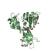
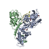
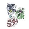
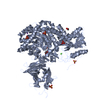
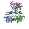
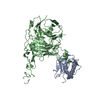
- PDB-1otc: THE O. NOVA TELOMERE END BINDING PROTEIN COMPLEXED WITH SINGLE ST... -
+ Open data

- Basic information
| Entry | Database: PDB / ID: 1otc | ||||||
|---|---|---|---|---|---|---|---|
| Title | THE O. NOVA TELOMERE END BINDING PROTEIN COMPLEXED WITH SINGLE STRAND DNA | ||||||
 Components Components |
| ||||||
 Keywords Keywords | PROTEIN/DNA / SINGLE STRAND DNA BINDING PROTEIN / PROTEIN DNA INTERACTIONS / PROTEIN PROTEIN INTERACTIONS / OLIGONUCLEOTIDE AND OLIGOSACCHARIDE BINDING FOLD / OB FOLD / TELOMERES / PROTEIN-DNA COMPLEX | ||||||
| Function / homology |  Function and homology information Function and homology informationtelomere cap complex / telomerase inhibitor activity / regulation of telomere maintenance via telomerase / single-stranded telomeric DNA binding / G-rich strand telomeric DNA binding / nuclear telomere cap complex / telomere capping / protein-containing complex / nucleus Similarity search - Function | ||||||
| Biological species |  Sterkiella nova (eukaryote) Sterkiella nova (eukaryote) | ||||||
| Method |  X-RAY DIFFRACTION / MIR / Resolution: 2.8 Å X-RAY DIFFRACTION / MIR / Resolution: 2.8 Å | ||||||
 Authors Authors | Horvath, M.P. / Schweiker, V.L. / Bevilacqua, J.M. / Ruggles, J.A. / Schultz, S.C. | ||||||
 Citation Citation | Journal: Cell(Cambridge,Mass.) / Year: 1998 Title: Crystal structure of the Oxytricha nova telomere end binding protein complexed with single strand DNA. Authors: Horvath, M.P. / Schweiker, V.L. / Bevilacqua, J.M. / Ruggles, J.A. / Schultz, S.C. | ||||||
| History |
|
- Structure visualization
| Structure viewer | Molecule: MolmilJmol/JSmol |
|---|
- Downloads & links
-Download
| PDBx/mmCIF format | 1otc.cif.gz | 151.5 KB | Display | PDBx/mmCIF format |
|---|---|---|---|---|
| PDB format | pdb1otc.ent.gz | 118 KB | Display | PDB format |
| PDBx/mmJSON format | 1otc.json.gz | Tree view | PDBx/mmJSON format | |
| Others |  Other downloads Other downloads |
-Validation report
| Arichive directory | https://data.pdbj.org/pub/pdb/validation_reports/ot/1otcftp://data.pdbj.org/pub/pdb/validation_reports/ot/1otc | HTTPS FTP |
|---|
-Related structure data
| Similar structure data |
|---|


-Links
 PDBj
PDBj


- Assembly
| Deposited unit | 
| ||||||||||
|---|---|---|---|---|---|---|---|---|---|---|---|
| 1 |
| ||||||||||
| Unit cell |
|
-Components
| #1: DNA chain | Mass: 3805.460 Da / Num. of mol.: 1 / Fragment: SINGLE STRAND DODECAMER DNA / Source method: obtained synthetically |
|---|---|
| #2: Protein | Mass: 56168.301 Da / Num. of mol.: 1 Source method: isolated from a genetically manipulated source Details: ALANINE VERSION / Source: (gene. exp.) Sterkiella nova (eukaryote) / Gene: MAC-56A / Organelle: MACRONUCLEOUS / Plasmid: PKKT7-H / Gene (production host): MAC-56A / Production host:  |
| #3: Protein | Mass: 28716.672 Da / Num. of mol.: 1 / Fragment: N-TERMINAL 28 KDA CORE DOMAIN Source method: isolated from a genetically manipulated source Details: ALANINE VERSION / Source: (gene. exp.) Sterkiella nova (eukaryote) / Gene: MAC-41A / Organelle: MACRONUCLEOUSPlasmid details: GENE WAS TRUNCTATED TO PRODUCE THE N-TERMINAL 28 KDA CORE DOMAIN ENDING WITH RESIDE LEU B 260 Plasmid: PKKT7-H / Gene (production host): MAC-41A / Production host: |
| #4: Water | ChemComp-HOH /  Mass: 18.015 Da / Num. of mol.: 23 / Source method: isolated from a natural source / Formula: H2O Mass: 18.015 Da / Num. of mol.: 23 / Source method: isolated from a natural source / Formula: H2O |
-Experimental details
-Experiment
| Experiment | Method: X-RAY DIFFRACTION / Number of used crystals: 2 |
|---|
- Sample preparation
| Crystal | Density Matthews: 3.4 Å3/Da / Density % sol: 60 % | |||||||||||||||||||||||||||||||||||||||||||||||||||||||||||||||||||||||||||||||||||||||||||||||||||||||||
|---|---|---|---|---|---|---|---|---|---|---|---|---|---|---|---|---|---|---|---|---|---|---|---|---|---|---|---|---|---|---|---|---|---|---|---|---|---|---|---|---|---|---|---|---|---|---|---|---|---|---|---|---|---|---|---|---|---|---|---|---|---|---|---|---|---|---|---|---|---|---|---|---|---|---|---|---|---|---|---|---|---|---|---|---|---|---|---|---|---|---|---|---|---|---|---|---|---|---|---|---|---|---|---|---|---|---|
| Crystal grow | Temperature: 293 K / Method: vapor diffusion, hanging drop / pH: 6 Details: pH 6.0, VAPOR DIFFUSION, HANGING DROP, temperature 293K | |||||||||||||||||||||||||||||||||||||||||||||||||||||||||||||||||||||||||||||||||||||||||||||||||||||||||
| Components of the solutions |
| |||||||||||||||||||||||||||||||||||||||||||||||||||||||||||||||||||||||||||||||||||||||||||||||||||||||||
| Crystal grow | *PLUS Details: drop contains the same amount of reservoir solution except 50mM NaCl | |||||||||||||||||||||||||||||||||||||||||||||||||||||||||||||||||||||||||||||||||||||||||||||||||||||||||
| Components of the solutions | *PLUS
|
-Data collection
| Diffraction | Mean temperature: 288 K |
|---|---|
| Diffraction source | Source: ROTATING ANODE / Type: RIGAKU RU200 / Wavelength: 1.5418 |
| Detector | Type: RIGAKU RAXIS IIC / Detector: IMAGE PLATE / Date: Aug 1, 1994 / Details: YALE MIRRORS |
| Radiation | Monochromator: NI FILTER / Protocol: SINGLE WAVELENGTH / Monochromatic (M) / Laue (L): M / Scattering type: x-ray |
| Radiation wavelength | Wavelength: 1.5418 Å / Relative weight: 1 |
| Reflection | Resolution: 2.8→30 Å / Num. obs: 24061 / % possible obs: 82.5 % / Redundancy: 2.9 % / Biso Wilson estimate: 6.9 Å2 / Rsym value: 0.084 / Net I/σ(I): 10.3 |
| Reflection shell | Resolution: 2.8→2.98 Å / Redundancy: 2.2 % / Mean I/σ(I) obs: 1.8 / Rsym value: 0.498 / % possible all: 57.1 |
| Reflection | *PLUS Num. measured all: 70514 / Rmerge(I) obs: 0.084 |
| Reflection shell | *PLUS % possible obs: 57.1 % / Rmerge(I) obs: 0.498 |
- Processing
| Software |
| ||||||||||||||||||||||||||||||||||||||||||||||||||||||||||||||||||||||||||||||||
|---|---|---|---|---|---|---|---|---|---|---|---|---|---|---|---|---|---|---|---|---|---|---|---|---|---|---|---|---|---|---|---|---|---|---|---|---|---|---|---|---|---|---|---|---|---|---|---|---|---|---|---|---|---|---|---|---|---|---|---|---|---|---|---|---|---|---|---|---|---|---|---|---|---|---|---|---|---|---|---|---|---|
| Refinement | Method to determine structure: MIR / Resolution: 2.8→20 Å / Rfactor Rfree error: 0.006 / Data cutoff high rms absF: 328629.15 / Isotropic thermal model: RESTRAINED / Cross valid method: THROUGHOUT / σ(F): 0
| ||||||||||||||||||||||||||||||||||||||||||||||||||||||||||||||||||||||||||||||||
| Solvent computation | Solvent model: FLAT MODEL / Bsol: 10 Å2 / ksol: 0.26 e/Å3 | ||||||||||||||||||||||||||||||||||||||||||||||||||||||||||||||||||||||||||||||||
| Displacement parameters | Biso mean: 37.1 Å2 | ||||||||||||||||||||||||||||||||||||||||||||||||||||||||||||||||||||||||||||||||
| Refine analyze |
| ||||||||||||||||||||||||||||||||||||||||||||||||||||||||||||||||||||||||||||||||
| Refinement step | Cycle: LAST / Resolution: 2.8→20 Å
| ||||||||||||||||||||||||||||||||||||||||||||||||||||||||||||||||||||||||||||||||
| Refine LS restraints |
| ||||||||||||||||||||||||||||||||||||||||||||||||||||||||||||||||||||||||||||||||
| LS refinement shell | Resolution: 2.8→2.97 Å / Rfactor Rfree error: 0.023 / Total num. of bins used: 6
| ||||||||||||||||||||||||||||||||||||||||||||||||||||||||||||||||||||||||||||||||
| Xplor file |
| ||||||||||||||||||||||||||||||||||||||||||||||||||||||||||||||||||||||||||||||||
| Software | *PLUS Name: CNS / Version: 0.5 / Classification: refinement | ||||||||||||||||||||||||||||||||||||||||||||||||||||||||||||||||||||||||||||||||
| Refinement | *PLUS | ||||||||||||||||||||||||||||||||||||||||||||||||||||||||||||||||||||||||||||||||
| Solvent computation | *PLUS | ||||||||||||||||||||||||||||||||||||||||||||||||||||||||||||||||||||||||||||||||
| Displacement parameters | *PLUS | ||||||||||||||||||||||||||||||||||||||||||||||||||||||||||||||||||||||||||||||||
| Refine LS restraints | *PLUS
| ||||||||||||||||||||||||||||||||||||||||||||||||||||||||||||||||||||||||||||||||
| LS refinement shell | *PLUS Rfactor obs: 0.361 |
