 Movie
Movie Controller

[English] 日本語
 Yorodumi
- PDB-2i0q: Crystal structure of a telomere single-strand DNA-protein complex... -
+ Open data

- Basic information
| Entry | Database: PDB / ID: 2i0q | ||||||
|---|---|---|---|---|---|---|---|
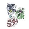
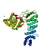
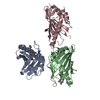
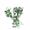
| Title | Crystal structure of a telomere single-strand DNA-protein complex from O. nova with full-length alpha and beta telomere proteins | ||||||
 Components Components |
| ||||||
 Keywords Keywords | structural protein/DNA / SINGLE STRAND DNA-PROTEIN COMPLEX / structural protein-DNA COMPLEX | ||||||
| Function / homology |  Function and homology information Function and homology informationtelomere cap complex / telomerase inhibitor activity / regulation of telomere maintenance via telomerase / single-stranded telomeric DNA binding / nuclear telomere cap complex / G-rich strand telomeric DNA binding / telomere capping / protein-containing complex / nucleus Similarity search - Function | ||||||
| Biological species |  Sterkiella nova (eukaryote) Sterkiella nova (eukaryote) | ||||||
| Method |  X-RAY DIFFRACTION / SYNCHROTRON / MOLECULAR REPLACEMENT / Resolution: 1.91 Å X-RAY DIFFRACTION / SYNCHROTRON / MOLECULAR REPLACEMENT / Resolution: 1.91 Å | ||||||
 Authors Authors | Horvath, M.P. / Buczek, P. | ||||||
 Citation Citation | Journal: J.Biol.Chem. / Year: 2006 Title: Structural reorganization and the cooperative binding of single-stranded telomere DNA in Sterkiella nova. Authors: Buczek, P. / Horvath, M.P. #1: Journal: Cell(Cambridge,Mass.) / Year: 1998Title: Crystal structure of the Oxytricha nova telomere end binding protein complexed with single strand DNA Authors: Horvath, M.P. / Schweiker, V.L. / Bevilacqua, J.M. / Ruggles, J.A. / Schultz, S.C. #2: Journal: J.Mol.Biol. / Year: 2001Title: DNA G-quartets in a 1.86 A resolution structure of an Oxytricha nova telomeric protein-DNA complex Authors: Horvath, M.P. / Schultz, S.C. | ||||||
| History |
|
- Structure visualization
| Structure viewer | Molecule: MolmilJmol/JSmol |
|---|
- Downloads & links
-Download
| PDBx/mmCIF format | 2i0q.cif.gz | 167 KB | Display | PDBx/mmCIF format |
|---|---|---|---|---|
| PDB format | pdb2i0q.ent.gz | 126.4 KB | Display | PDB format |
| PDBx/mmJSON format | 2i0q.json.gz | Tree view | PDBx/mmJSON format | |
| Others |  Other downloads Other downloads |
-Validation report
| Arichive directory | https://data.pdbj.org/pub/pdb/validation_reports/i0/2i0qftp://data.pdbj.org/pub/pdb/validation_reports/i0/2i0q | HTTPS FTP |
|---|
-Related structure data
| Related structure data |  1jb7S S: Starting model for refinement |
|---|---|
| Similar structure data |


-Links
 PDBj
PDBj


- Assembly
| Deposited unit | 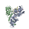
| ||||||||
|---|---|---|---|---|---|---|---|---|---|
| 1 |
| ||||||||
| Unit cell |
| ||||||||
| Components on special symmetry positions |
|
-Components
-DNA chain , 1 types, 1 molecules D
| #1: DNA chain | Mass: 3476.254 Da / Num. of mol.: 1 / Source method: obtained synthetically Details: 3'-terminal single strand DNA fragment from Oxytricha nova (Sterkiella nova) macronuclear telomeres |
|---|
-Telomere-binding protein ... , 2 types, 2 molecules AB
| #2: Protein | Mass: 56168.301 Da / Num. of mol.: 1 Source method: isolated from a genetically manipulated source Source: (gene. exp.) Sterkiella nova (eukaryote) / Gene: MAC-56A / Plasmid: pKKT7e / Production host:  |
|---|---|
| #3: Protein | Mass: 41527.527 Da / Num. of mol.: 1 Source method: isolated from a genetically manipulated source Source: (gene. exp.) Sterkiella nova (eukaryote) / Gene: MAC-41A / Plasmid: pKKT7e / Production host: |
-Non-polymers , 3 types, 315 molecules 


| #4: Chemical | ChemComp-CL /  Mass: 35.453 Da / Num. of mol.: 4 / Source method: obtained synthetically / Formula: Cl Mass: 35.453 Da / Num. of mol.: 4 / Source method: obtained synthetically / Formula: Cl#5: Chemical | ChemComp-EDO /  Mass: 62.068 Da / Num. of mol.: 5 / Source method: obtained synthetically / Formula: C2H6O2 Mass: 62.068 Da / Num. of mol.: 5 / Source method: obtained synthetically / Formula: C2H6O2#6: Water | ChemComp-HOH / | Mass: 18.015 Da / Num. of mol.: 306 / Source method: isolated from a natural source / Formula: H2O |
|---|
-Experimental details
-Experiment
| Experiment | Method: X-RAY DIFFRACTION / Number of used crystals: 1 |
|---|
- Sample preparation
| Crystal | Density Matthews: 2.63 Å3/Da / Density % sol: 53.23 % | ||||||||||||||||||||||||||||||||||||||||||||||||
|---|---|---|---|---|---|---|---|---|---|---|---|---|---|---|---|---|---|---|---|---|---|---|---|---|---|---|---|---|---|---|---|---|---|---|---|---|---|---|---|---|---|---|---|---|---|---|---|---|---|
| Crystal grow | Temperature: 277.15 K / Method: vapor diffusion, hanging drop / pH: 5.75 Details: 21% (w/v) PEG-4000, 10% (v/v) ethylene glycol, 1.5 M sodium chloride, 40 mM MES, 2 mM DTT, 0.02% sodium azide, VAPOR DIFFUSION, HANGING DROP, pH 5.75, temperature 277.15K | ||||||||||||||||||||||||||||||||||||||||||||||||
| Components of the solutions |
|
-Data collection
| Diffraction | Mean temperature: 100 K |
|---|---|
| Diffraction source | Source: SYNCHROTRON / Site: ALS  / Beamline: 12.3.1 / Wavelength: 1.11587 Å / Beamline: 12.3.1 / Wavelength: 1.11587 Å |
| Detector | Type: ADSC QUANTUM 315 / Detector: CCD / Date: Apr 1, 2006 |
| Radiation | Protocol: SINGLE WAVELENGTH / Monochromatic (M) / Laue (L): M / Scattering type: x-ray |
| Radiation wavelength | Wavelength: 1.11587 Å / Relative weight: 1 |
| Reflection | Resolution: 1.91→50 Å / Num. all: 86086 / Num. obs: 85120 / % possible obs: 98.9 % / Observed criterion σ(F): 0 / Observed criterion σ(I): -3 / Redundancy: 8.1 % / Biso Wilson estimate: 47.6 Å2 / Rsym value: 0.041 / Net I/σ(I): 38.8 |
| Reflection shell | Resolution: 1.91→1.98 Å / Redundancy: 5.3 % / Mean I/σ(I) obs: 3.5 / Rsym value: 0.46 / % possible all: 95.7 |
- Processing
| Software |
| |||||||||||||||||||||||||||||||||||||||||||||||||
|---|---|---|---|---|---|---|---|---|---|---|---|---|---|---|---|---|---|---|---|---|---|---|---|---|---|---|---|---|---|---|---|---|---|---|---|---|---|---|---|---|---|---|---|---|---|---|---|---|---|---|
| Refinement | Method to determine structure: MOLECULAR REPLACEMENT Starting model: PDB ENTRY 1JB7 Resolution: 1.91→50 Å / Isotropic thermal model: Isotropic / Cross valid method: THROUGHOUT / σ(F): 0 / σ(I): -3 / Stereochemistry target values: Engh & Huber Details: Maximum likelihood refinement with intensities as target function (mli)
| |||||||||||||||||||||||||||||||||||||||||||||||||
| Solvent computation | Solvent model: CNS SOLVE / Bsol: 30.2205 Å2 / ksol: 0.320563 e/Å3 | |||||||||||||||||||||||||||||||||||||||||||||||||
| Displacement parameters | Biso mean: 43.3 Å2 | |||||||||||||||||||||||||||||||||||||||||||||||||
| Refine analyze |
| |||||||||||||||||||||||||||||||||||||||||||||||||
| Refinement step | Cycle: LAST / Resolution: 1.91→50 Å
| |||||||||||||||||||||||||||||||||||||||||||||||||
| Refine LS restraints |
| |||||||||||||||||||||||||||||||||||||||||||||||||
| LS refinement shell | Refine-ID: X-RAY DIFFRACTION
|
