 Movie
Movie Controller

[English] 日本語
 Yorodumi
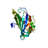
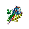
- PDB-5ys0: Crystal structure of the second StARkin domain of Lam2 in complex... -
+ Open data

- Basic information
| Entry | Database: PDB / ID: 5ys0 | ||||||
|---|---|---|---|---|---|---|---|
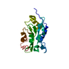
| Title | Crystal structure of the second StARkin domain of Lam2 in complex with ergosterol | ||||||
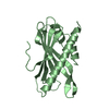
 Components Components | Membrane-anchored lipid-binding protein YSP2 | ||||||
 Keywords Keywords | TRANSPORT PROTEIN / ligand binding domain / sterol / lipid transport / Ysp2 | ||||||
| Function / homology |  Function and homology information Function and homology informationintracellular sterol transport / endoplasmic reticulum-plasma membrane contact site / sterol transfer activity / sterol binding / sterol transport / cortical endoplasmic reticulum / cell periphery / mitochondrial membrane / apoptotic process / endoplasmic reticulum membrane ...intracellular sterol transport / endoplasmic reticulum-plasma membrane contact site / sterol transfer activity / sterol binding / sterol transport / cortical endoplasmic reticulum / cell periphery / mitochondrial membrane / apoptotic process / endoplasmic reticulum membrane / mitochondrion / plasma membrane / cytoplasm Similarity search - Function | ||||||
| Biological species |  | ||||||
| Method |  X-RAY DIFFRACTION / SYNCHROTRON / MOLECULAR REPLACEMENT / Resolution: 2.601 Å X-RAY DIFFRACTION / SYNCHROTRON / MOLECULAR REPLACEMENT / Resolution: 2.601 Å | ||||||
 Authors Authors | Tong, J. / Im, Y.J. | ||||||
| Funding support |  Korea, Republic Of, 1items Korea, Republic Of, 1items
| ||||||
 Citation Citation | Journal: Proc. Natl. Acad. Sci. U.S.A. / Year: 2018 Title: Structural basis of sterol recognition and nonvesicular transport by lipid transfer proteins anchored at membrane contact sites Authors: Tong, J. / Manik, M.K. / Im, Y.J. | ||||||
| History |
|
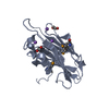
- Structure visualization
| Structure viewer | Molecule: MolmilJmol/JSmol |
|---|
- Downloads & links
-Download
| PDBx/mmCIF format | 5ys0.cif.gz | 108.5 KB | Display | PDBx/mmCIF format |
|---|---|---|---|---|
| PDB format | pdb5ys0.ent.gz | 82.6 KB | Display | PDB format |
| PDBx/mmJSON format | 5ys0.json.gz | Tree view | PDBx/mmJSON format | |
| Others |  Other downloads Other downloads |
-Validation report
| Arichive directory | https://data.pdbj.org/pub/pdb/validation_reports/ys/5ys0ftp://data.pdbj.org/pub/pdb/validation_reports/ys/5ys0 | HTTPS FTP |
|---|
-Related structure data
| Related structure data |  5yqiC  5yqjC 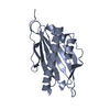 5yqpC 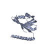 5yqqC 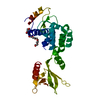 5yqrC 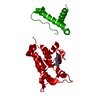 4yqjS S: Starting model for refinement C: citing same article ( |
|---|---|
| Similar structure data |


-Links
 PDBj
PDBj- Assembly
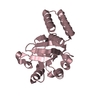
| Deposited unit | 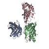
| ||||||||
|---|---|---|---|---|---|---|---|---|---|
| 1 | 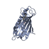
| ||||||||
| 2 | 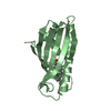
| ||||||||
| 3 | 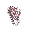
| ||||||||
| Unit cell |
|
-Components
| #1: Protein | Mass: 19133.781 Da / Num. of mol.: 3 Source method: isolated from a genetically manipulated source Source: (gene. exp.) Strain: ATCC 204508 / S288c / Gene: YSP2, LAM2, LTC4, YDR326C / Plasmid: pHIS2-Thr Details (production host): N-terminal hexahistidine tag fusion Production host:  #2: Chemical |   Mass: 396.648 Da / Num. of mol.: 2 / Source method: obtained synthetically / Formula: C28H44O / Feature type: SUBJECT OF INVESTIGATION Mass: 396.648 Da / Num. of mol.: 2 / Source method: obtained synthetically / Formula: C28H44O / Feature type: SUBJECT OF INVESTIGATION#3: Water | ChemComp-HOH / |  Mass: 18.015 Da / Num. of mol.: 21 / Source method: isolated from a natural source / Formula: H2O Mass: 18.015 Da / Num. of mol.: 21 / Source method: isolated from a natural source / Formula: H2O |
|---|
-Experimental details
-Experiment
| Experiment | Method: X-RAY DIFFRACTION / Number of used crystals: 1 |
|---|
- Sample preparation
| Crystal | Density Matthews: 2.41 Å3/Da / Density % sol: 48.86 % |
|---|---|
| Crystal grow | Temperature: 295 K / Method: vapor diffusion, hanging drop / pH: 8 / Details: 0.1M Tris-HCl pH 8.0, 30% PEG8000. |
-Data collection
| Diffraction | Mean temperature: 100 K |
|---|---|
| Diffraction source | Source: SYNCHROTRON / Site: PAL/PLS / Beamline: 7A (6B, 6C1) / Wavelength: 0.9795 Å |
| Detector | Type: ADSC QUANTUM 270 / Detector: CCD / Date: Dec 13, 2015 |
| Radiation | Protocol: SINGLE WAVELENGTH / Monochromatic (M) / Laue (L): M / Scattering type: x-ray |
| Radiation wavelength | Wavelength: 0.9795 Å / Relative weight: 1 |
| Reflection | Resolution: 2.6→50 Å / Num. obs: 16717 / % possible obs: 99 % / Redundancy: 4.8 % / Biso Wilson estimate: 63.5 Å2 / Rmerge(I) obs: 0.039 / Net I/σ(I): 48.9 |
| Reflection shell | Resolution: 2.6→2.64 Å / Redundancy: 4.8 % / Rmerge(I) obs: 0.331 / Mean I/σ(I) obs: 6.9 / Num. unique obs: 845 / % possible all: 99.8 |
- Processing
| Software |
| |||||||||||||||||||||||||||||||||||||||||||||||||
|---|---|---|---|---|---|---|---|---|---|---|---|---|---|---|---|---|---|---|---|---|---|---|---|---|---|---|---|---|---|---|---|---|---|---|---|---|---|---|---|---|---|---|---|---|---|---|---|---|---|---|
| Refinement | Method to determine structure: MOLECULAR REPLACEMENT Starting model: 4YQJ Resolution: 2.601→32.709 Å / SU ML: 0.33 / Cross valid method: FREE R-VALUE / σ(F): 1.34 / Phase error: 33.29
| |||||||||||||||||||||||||||||||||||||||||||||||||
| Solvent computation | Shrinkage radii: 0.9 Å / VDW probe radii: 1.11 Å | |||||||||||||||||||||||||||||||||||||||||||||||||
| Refinement step | Cycle: LAST / Resolution: 2.601→32.709 Å
| |||||||||||||||||||||||||||||||||||||||||||||||||
| Refine LS restraints |
| |||||||||||||||||||||||||||||||||||||||||||||||||
| LS refinement shell |
|
