 Movie
Movie Controller

[English] 日本語
 Yorodumi
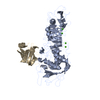
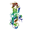
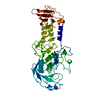
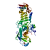
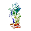
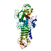
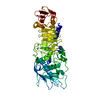
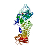
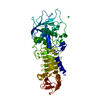
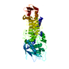
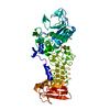
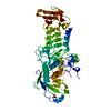
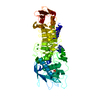
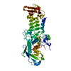
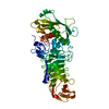
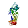
- PDB-1o0t: CRYSTAL STRUCTURE OF A COLD ADAPTED ALKALINE PROTEASE FROM PSEUDO... -
+ Open data

- Basic information
| Entry | Database: PDB / ID: 1o0t | ||||||
|---|---|---|---|---|---|---|---|
| Title | CRYSTAL STRUCTURE OF A COLD ADAPTED ALKALINE PROTEASE FROM PSEUDOMONAS TAC II 18, CO-CRYSTALLIZED WITH 5 mM EDTA (5 DAYS) | ||||||
 Components Components | serralysin | ||||||
 Keywords Keywords | HYDROLASE / BETA JELLY ROLL / CALCIUM AND ZINC DEPENDENT ALKALINE PROTEASE | ||||||
| Function / homology |  Function and homology information Function and homology informationserralysin / metalloendopeptidase activity / extracellular matrix / calcium ion binding / proteolysis / : / zinc ion binding Similarity search - Function | ||||||
| Biological species |  Pseudomonas sp. 'TAC II 18' (bacteria) Pseudomonas sp. 'TAC II 18' (bacteria) | ||||||
| Method |  X-RAY DIFFRACTION / MOLECULAR REPLACEMENT / Resolution: 2.5 Å X-RAY DIFFRACTION / MOLECULAR REPLACEMENT / Resolution: 2.5 Å | ||||||
 Authors Authors | Ravaud, S. / Gouet, P. / Haser, R. / Aghajari, N. | ||||||
 Citation Citation | Journal: J.Bacteriol. / Year: 2003 Title: Probing the role of divalent metal ions in a bacterial psychrophilic metalloprotease: binding studies of an enzyme in the crystalline state by x-ray crystallography. Authors: Ravaud, S. / Gouet, P. / Haser, R. / Aghajari, N. | ||||||
| History |
|
- Structure visualization
| Structure viewer | Molecule: MolmilJmol/JSmol |
|---|
- Downloads & links
-Download
| PDBx/mmCIF format | 1o0t.cif.gz | 103.2 KB | Display | PDBx/mmCIF format |
|---|---|---|---|---|
| PDB format | pdb1o0t.ent.gz | 77.5 KB | Display | PDB format |
| PDBx/mmJSON format | 1o0t.json.gz | Tree view | PDBx/mmJSON format | |
| Others |  Other downloads Other downloads |
-Validation report
| Arichive directory | https://data.pdbj.org/pub/pdb/validation_reports/o0/1o0tftp://data.pdbj.org/pub/pdb/validation_reports/o0/1o0t | HTTPS FTP |
|---|
-Related structure data
| Related structure data |  1o0qC  1om6C  1om7C  1om8C  1omjC  1g9kS S: Starting model for refinement C: citing same article ( |
|---|---|
| Similar structure data |


-Links
 PDBj
PDBj


- Assembly
| Deposited unit | 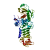
| ||||||||
|---|---|---|---|---|---|---|---|---|---|
| 1 |
| ||||||||
| 2 | 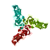
| ||||||||
| Unit cell |
|
-Components
| #1: Protein | Mass: 48727.609 Da / Num. of mol.: 1 / Source method: isolated from a natural source / Source: (natural) Pseudomonas sp. 'TAC II 18' (bacteria) / Strain: TAC II 18 / References: UniProt: O69771, serralysin | ||||
|---|---|---|---|---|---|
| #2: Chemical | ChemComp-CA /   Mass: 40.078 Da / Num. of mol.: 7 / Source method: obtained synthetically / Formula: Ca Mass: 40.078 Da / Num. of mol.: 7 / Source method: obtained synthetically / Formula: Ca#3: Chemical | ChemComp-SO4 / |   Mass: 96.063 Da / Num. of mol.: 1 / Source method: obtained synthetically / Formula: SO4 Mass: 96.063 Da / Num. of mol.: 1 / Source method: obtained synthetically / Formula: SO4#4: Water | ChemComp-HOH / |  Mass: 18.015 Da / Num. of mol.: 161 / Source method: isolated from a natural source / Formula: H2O Mass: 18.015 Da / Num. of mol.: 161 / Source method: isolated from a natural source / Formula: H2O |
-Experimental details
-Experiment
| Experiment | Method: X-RAY DIFFRACTION / Number of used crystals: 1 |
|---|
- Sample preparation
| Crystal | Density Matthews: 2.55 Å3/Da / Density % sol: 51.41 % | ||||||||||||||||||||||||
|---|---|---|---|---|---|---|---|---|---|---|---|---|---|---|---|---|---|---|---|---|---|---|---|---|---|
| Crystal grow | Temperature: 289 K / Method: vapor diffusion, hanging drop / pH: 7 Details: Ammonium sulfate, Hepes, pH 7.00, VAPOR DIFFUSION, HANGING DROP, temperature 289K | ||||||||||||||||||||||||
| Crystal grow | *PLUS Temperature: 4 ℃ / pH: 7 / Method: vapor diffusion, hanging drop / Details: Villeret, V., (1997) Protein Sci., 6, 2462. | ||||||||||||||||||||||||
| Components of the solutions | *PLUS
|
-Data collection
| Diffraction | Mean temperature: 288 K |
|---|---|
| Diffraction source | Source: ROTATING ANODE / Type: ENRAF-NONIUS FR591 / Wavelength: 1.5418 / Wavelength: 1.5418 Å |
| Detector | Type: MARRESEARCH / Detector: IMAGE PLATE / Date: Feb 5, 2002 / Details: MIRRORS |
| Radiation | Monochromator: OSMIC MIRRORS / Protocol: SINGLE WAVELENGTH / Monochromatic (M) / Laue (L): M / Scattering type: x-ray |
| Radiation wavelength | Wavelength: 1.5418 Å / Relative weight: 1 |
| Reflection | Resolution: 2.5→32.3 Å / Num. all: 16749 / Num. obs: 16749 / % possible obs: 99.1 % / Observed criterion σ(F): 2 / Observed criterion σ(I): 2 / Redundancy: 2 % / Rsym value: 0.116 |
| Reflection shell | Resolution: 2.5→2.6 Å / Redundancy: 2 % / Rsym value: 0.374 / % possible all: 98.3 |
| Reflection | *PLUS Num. measured all: 33198 / Rmerge(I) obs: 0.116 |
| Reflection shell | *PLUS % possible obs: 98.3 % / Rmerge(I) obs: 0.374 / Mean I/σ(I) obs: 1.8 |
- Processing
| Software |
| ||||||||||||||||||||
|---|---|---|---|---|---|---|---|---|---|---|---|---|---|---|---|---|---|---|---|---|---|
| Refinement | Method to determine structure: MOLECULAR REPLACEMENT Starting model: PDB ENTRY 1G9K Resolution: 2.5→50 Å / Cross valid method: THROUGHOUT / σ(F): 2 / σ(I): 2 / Stereochemistry target values: ENGH & HUBER
| ||||||||||||||||||||
| Refinement step | Cycle: LAST / Resolution: 2.5→50 Å
| ||||||||||||||||||||
| Refine LS restraints |
|
