 Movie
Movie Controller


+ Open data

- Basic information
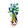
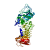
| Entry | Database: PDB / ID: 1akl | ||||||
|---|---|---|---|---|---|---|---|
| Title | ALKALINE PROTEASE FROM PSEUDOMONAS AERUGINOSA IFO3080 | ||||||
 Components Components | ALKALINE PROTEASE | ||||||
 Keywords Keywords | HYDROLASE (METALLOPROTEINASE) | ||||||
| Function / homology |  Function and homology information Function and homology informationsymbiont-mediated evasion of recognition by host pattern recognition receptor / serralysin / negative regulation of complement activation, lectin pathway / negative regulation of complement activation, classical pathway / positive regulation of sodium ion transport / metalloendopeptidase activity / peptidase activity / calcium ion binding / proteolysis / : / zinc ion binding Similarity search - Function | ||||||
| Biological species |   Pseudomonas aeruginosa (bacteria) Pseudomonas aeruginosa (bacteria) | ||||||
| Method |  X-RAY DIFFRACTION / SYNCHROTRON / Resolution: 2 Å X-RAY DIFFRACTION / SYNCHROTRON / Resolution: 2 Å | ||||||
 Authors Authors | Miyatake, H. / Hata, Y. / Fujii, T. / Hamada, K. / Morihara, K. / Katsube, Y. | ||||||
 Citation Citation | Journal: J.Biochem.(Tokyo) / Year: 1995 Title: Crystal structure of the unliganded alkaline protease from Pseudomonas aeruginosa IFO3080 and its conformational changes on ligand binding. Authors: Miyatake, H. / Hata, Y. / Fujii, T. / Hamada, K. / Morihara, K. / Katsube, Y. #1: Journal: Photon Factory Activity Report / Year: 1993Title: Crystal Structure Determination of Alkaline Protease from Pseudomonas Aeruginosa Strain Pao1 Authors: Miyatake, H. / Hata, Y. / Fujii, T. / Morihara, K. / Katsube, Y. | ||||||
| History |
|
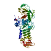
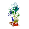
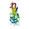
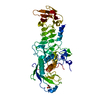
- Structure visualization
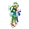
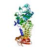
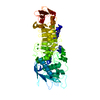
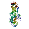
| Structure viewer | Molecule: MolmilJmol/JSmol |
|---|
- Downloads & links
-Download
| PDBx/mmCIF format | 1akl.cif.gz | 108.8 KB | Display | PDBx/mmCIF format |
|---|---|---|---|---|
| PDB format | pdb1akl.ent.gz | 82.4 KB | Display | PDB format |
| PDBx/mmJSON format | 1akl.json.gz | Tree view | PDBx/mmJSON format | |
| Others |  Other downloads Other downloads |
-Validation report
| Arichive directory | https://data.pdbj.org/pub/pdb/validation_reports/ak/1aklftp://data.pdbj.org/pub/pdb/validation_reports/ak/1akl | HTTPS FTP |
|---|
-Related structure data
| Similar structure data |
|---|

-Links
 PDBj
PDBj


- Assembly
| Deposited unit | 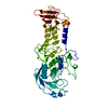
| ||||||||
|---|---|---|---|---|---|---|---|---|---|
| 1 |
| ||||||||
| Unit cell |
|
-Components
| #1: Protein | Mass: 49531.426 Da / Num. of mol.: 1 / Source method: isolated from a natural source / Source: (natural) Pseudomonas aeruginosa (bacteria) / Strain: IFO3080 / References: UniProt: Q03023, serralysin | ||
|---|---|---|---|
| #2: Chemical | ChemComp-ZN /   Mass: 65.409 Da / Num. of mol.: 1 / Source method: obtained synthetically / Formula: Zn Mass: 65.409 Da / Num. of mol.: 1 / Source method: obtained synthetically / Formula: Zn | ||
| #3: Chemical | ChemComp-CA /   Mass: 40.078 Da / Num. of mol.: 8 / Source method: obtained synthetically / Formula: Ca Mass: 40.078 Da / Num. of mol.: 8 / Source method: obtained synthetically / Formula: Ca#4: Water | ChemComp-HOH / |  Mass: 18.015 Da / Num. of mol.: 331 / Source method: isolated from a natural source / Formula: H2O Mass: 18.015 Da / Num. of mol.: 331 / Source method: isolated from a natural source / Formula: H2O |
-Experimental details
-Experiment
| Experiment | Method: X-RAY DIFFRACTION |
|---|
- Sample preparation
| Crystal | Density Matthews: 3.52 Å3/Da / Density % sol: 65.03 % | ||||||||||||||||||||||||||||||||||||||||||||||||||||||
|---|---|---|---|---|---|---|---|---|---|---|---|---|---|---|---|---|---|---|---|---|---|---|---|---|---|---|---|---|---|---|---|---|---|---|---|---|---|---|---|---|---|---|---|---|---|---|---|---|---|---|---|---|---|---|---|
| Crystal | *PLUS Density % sol: 66 % | ||||||||||||||||||||||||||||||||||||||||||||||||||||||
| Crystal grow | *PLUS Temperature: 25 K / pH: 7 / Method: vapor diffusion, hanging drop | ||||||||||||||||||||||||||||||||||||||||||||||||||||||
| Components of the solutions | *PLUS
|
-Data collection
| Diffraction source | Source: SYNCHROTRON / Site: Photon Factory  / Beamline: BL-6A / Wavelength: 1 / Beamline: BL-6A / Wavelength: 1 |
|---|---|
| Detector | Type: MARRESEARCH / Detector: IMAGE PLATE / Date: May 25, 1993 |
| Radiation | Monochromatic (M) / Laue (L): M / Scattering type: x-ray |
| Radiation wavelength | Wavelength: 1 Å / Relative weight: 1 |
| Reflection | Resolution: 2→50 Å / Num. obs: 44289 / Observed criterion σ(I): 1 / Rmerge(I) obs: 0.058 |
| Reflection | *PLUS Rmerge(I) obs: 0.058 |
- Processing
| Software |
| ||||||||||||||||||||||||||||||||||||||||||||||||||||||||||||
|---|---|---|---|---|---|---|---|---|---|---|---|---|---|---|---|---|---|---|---|---|---|---|---|---|---|---|---|---|---|---|---|---|---|---|---|---|---|---|---|---|---|---|---|---|---|---|---|---|---|---|---|---|---|---|---|---|---|---|---|---|---|
| Refinement | Resolution: 2→8 Å / σ(F): 2
| ||||||||||||||||||||||||||||||||||||||||||||||||||||||||||||
| Displacement parameters | Biso mean: 38.12 Å2 | ||||||||||||||||||||||||||||||||||||||||||||||||||||||||||||
| Refine analyze | Luzzati coordinate error obs: 0.3 Å | ||||||||||||||||||||||||||||||||||||||||||||||||||||||||||||
| Refinement step | Cycle: LAST / Resolution: 2→8 Å
| ||||||||||||||||||||||||||||||||||||||||||||||||||||||||||||
| Refine LS restraints |
| ||||||||||||||||||||||||||||||||||||||||||||||||||||||||||||
| Software | *PLUS Name: X-PLOR / Classification: refinement | ||||||||||||||||||||||||||||||||||||||||||||||||||||||||||||
| Refinement | *PLUS | ||||||||||||||||||||||||||||||||||||||||||||||||||||||||||||
| Solvent computation | *PLUS | ||||||||||||||||||||||||||||||||||||||||||||||||||||||||||||
| Displacement parameters | *PLUS | ||||||||||||||||||||||||||||||||||||||||||||||||||||||||||||
| Refine LS restraints | *PLUS
|
