 Movie
Movie Controller

[English] 日本語
 Yorodumi
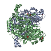
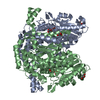
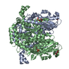
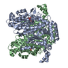
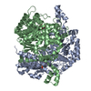
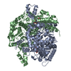
- PDB-1g98: CRYSTAL STRUCTURE ANALYSIS OF RABBIT PHOSPHOGLUCOSE ISOMERASE COM... -
+ Open data

- Basic information
| Entry | Database: PDB / ID: 1g98 | ||||||
|---|---|---|---|---|---|---|---|
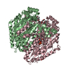
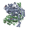
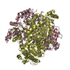
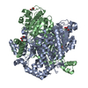
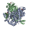
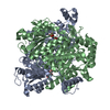
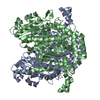
| Title | CRYSTAL STRUCTURE ANALYSIS OF RABBIT PHOSPHOGLUCOSE ISOMERASE COMPLEXED WITH 5-PHOSPHOARABINONATE, A TRANSITION STATE ANALOGUE | ||||||
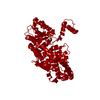
 Components Components | PHOSPHOGLUCOSE ISOMERASE | ||||||
 Keywords Keywords | ISOMERASE / Phosphoglucose Isomerase / 5-phosphoarabinonate / transition state analogue | ||||||
| Function / homology |  Function and homology information Function and homology informationIsomerases; Racemases and epimerases; Acting on carbohydrates and derivatives / racemase and epimerase activity, acting on carbohydrates and derivatives / glucose-6-phosphate isomerase / glucose-6-phosphate isomerase activity / glucose 6-phosphate metabolic process / carbohydrate derivative binding / monosaccharide binding / cytokine activity / gluconeogenesis / glycolytic process ...Isomerases; Racemases and epimerases; Acting on carbohydrates and derivatives / racemase and epimerase activity, acting on carbohydrates and derivatives / glucose-6-phosphate isomerase / glucose-6-phosphate isomerase activity / glucose 6-phosphate metabolic process / carbohydrate derivative binding / monosaccharide binding / cytokine activity / gluconeogenesis / glycolytic process / : / cytosol Similarity search - Function | ||||||
| Biological species |  | ||||||
| Method |  X-RAY DIFFRACTION / SYNCHROTRON / MOLECULAR REPLACEMENT / Resolution: 1.9 Å X-RAY DIFFRACTION / SYNCHROTRON / MOLECULAR REPLACEMENT / Resolution: 1.9 Å | ||||||
 Authors Authors | Jeffery, C.J. / Hardre, R. / Salmon, L. | ||||||
 Citation Citation | Journal: Biochemistry / Year: 2001 Title: Crystal structure of rabbit phosphoglucose isomerase complexed with 5-phospho-D-arabinonate identifies the role of Glu357 in catalysis. Authors: Jeffery, C.J. / Hardre, R. / Salmon, L. | ||||||
| History |
|
- Structure visualization
| Structure viewer | Molecule: MolmilJmol/JSmol |
|---|
- Downloads & links
-Download
| PDBx/mmCIF format | 1g98.cif.gz | 247 KB | Display | PDBx/mmCIF format |
|---|---|---|---|---|
| PDB format | pdb1g98.ent.gz | 196.6 KB | Display | PDB format |
| PDBx/mmJSON format | 1g98.json.gz | Tree view | PDBx/mmJSON format | |
| Others |  Other downloads Other downloads |
-Validation report
| Arichive directory | https://data.pdbj.org/pub/pdb/validation_reports/g9/1g98ftp://data.pdbj.org/pub/pdb/validation_reports/g9/1g98 | HTTPS FTP |
|---|
-Related structure data
| Related structure data |  1dqrS S: Starting model for refinement |
|---|---|
| Similar structure data |


-Links
 PDBj
PDBj

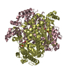
- Assembly
| Deposited unit | 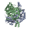
| ||||||||||
|---|---|---|---|---|---|---|---|---|---|---|---|
| 1 |
| ||||||||||
| Unit cell |
| ||||||||||
| Components on special symmetry positions |
|
-Components
| #1: Protein | Mass: 62827.621 Da / Num. of mol.: 2 / Source method: isolated from a natural source / Details: RABBIT SKELETAL MUSCLE TISSUE / Source: (natural) #2: Sugar | 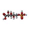  Type: saccharide / Mass: 246.109 Da / Num. of mol.: 2 / Source method: obtained synthetically / Formula: C5H11O9P Type: saccharide / Mass: 246.109 Da / Num. of mol.: 2 / Source method: obtained synthetically / Formula: C5H11O9P#3: Water | ChemComp-HOH / |  Mass: 18.015 Da / Num. of mol.: 892 / Source method: isolated from a natural source / Formula: H2O Mass: 18.015 Da / Num. of mol.: 892 / Source method: isolated from a natural source / Formula: H2O |
|---|
-Experimental details
-Experiment
| Experiment | Method: X-RAY DIFFRACTION / Number of used crystals: 1 |
|---|
- Sample preparation
| Crystal | Density Matthews: 2.61 Å3/Da / Density % sol: 52.87 % | ||||||||||||||||||||||||||||||||||||||||||||||||||||||
|---|---|---|---|---|---|---|---|---|---|---|---|---|---|---|---|---|---|---|---|---|---|---|---|---|---|---|---|---|---|---|---|---|---|---|---|---|---|---|---|---|---|---|---|---|---|---|---|---|---|---|---|---|---|---|---|
| Crystal grow | Temperature: 298 K / Method: vapor diffusion, hanging drop / pH: 7 Details: protein solution: 15-20mg/ml PGI, 10mM imidazole (pH 7.5), 50mM KCl, 3mMNaN3, 5mM 5PAH reservoir solution: 10-15% PEG 8000, 250mM magnesium acetate, 100mM sodium cacodylate (pH6.5). VAPOR ...Details: protein solution: 15-20mg/ml PGI, 10mM imidazole (pH 7.5), 50mM KCl, 3mMNaN3, 5mM 5PAH reservoir solution: 10-15% PEG 8000, 250mM magnesium acetate, 100mM sodium cacodylate (pH6.5). VAPOR DIFFUSION, HANGING DROP at 298K, pH 7.0, temperature 298.0K | ||||||||||||||||||||||||||||||||||||||||||||||||||||||
| Crystal grow | *PLUS pH: 7.5 | ||||||||||||||||||||||||||||||||||||||||||||||||||||||
| Components of the solutions | *PLUS
|
-Data collection
| Diffraction | Mean temperature: 93 K |
|---|---|
| Diffraction source | Source: SYNCHROTRON / Site: APS  / Beamline: 14-BM-C / Wavelength: 1 Å / Beamline: 14-BM-C / Wavelength: 1 Å |
| Detector | Type: ADSC QUANTUM 4 / Detector: CCD / Date: Nov 22, 1999 |
| Radiation | Protocol: SINGLE WAVELENGTH / Monochromatic (M) / Laue (L): M / Scattering type: x-ray |
| Radiation wavelength | Wavelength: 1 Å / Relative weight: 1 |
| Reflection | Resolution: 1.9→8 Å / Num. all: 95997 / Num. obs: 95997 / % possible obs: 94.3 % / Observed criterion σ(F): 0 / Observed criterion σ(I): 0 / Redundancy: 5.1 % / Biso Wilson estimate: 22.646 Å2 / Rmerge(I) obs: 0.035 / Rsym value: 0.035 / Net I/σ(I): 26.86 |
| Reflection shell | Resolution: 1.9→1.97 Å / Redundancy: 4.88 % / Rmerge(I) obs: 0.082 / Mean I/σ(I) obs: 15.45 / Num. unique all: 10080 / Rsym value: 0.082 / % possible all: 99.8 |
| Reflection | *PLUS Num. measured all: 486988 |
| Reflection shell | *PLUS % possible obs: 99.8 % |
- Processing
| Software |
| |||||||||||||||||||||||||
|---|---|---|---|---|---|---|---|---|---|---|---|---|---|---|---|---|---|---|---|---|---|---|---|---|---|---|
| Refinement | Method to determine structure: MOLECULAR REPLACEMENT Starting model: 1DQR Resolution: 1.9→8 Å / Isotropic thermal model: anisotropic / Cross valid method: THROUGHOUT / σ(F): 0 / σ(I): 0 / Stereochemistry target values: CNS
| |||||||||||||||||||||||||
| Displacement parameters | Biso mean: 25.96 Å2 | |||||||||||||||||||||||||
| Refine analyze |
| |||||||||||||||||||||||||
| Refinement step | Cycle: LAST / Resolution: 1.9→8 Å
| |||||||||||||||||||||||||
| Refine LS restraints |
| |||||||||||||||||||||||||
| LS refinement shell | Resolution: 1.9→1.97 Å
| |||||||||||||||||||||||||
| Software | *PLUS Name: CNS / Classification: refinement | |||||||||||||||||||||||||
| Refinement | *PLUS Highest resolution: 1.9 Å / Lowest resolution: 8 Å / σ(F): 0 / % reflection Rfree: 10 % / Rfactor obs: 0.211 | |||||||||||||||||||||||||
| Solvent computation | *PLUS | |||||||||||||||||||||||||
| Displacement parameters | *PLUS | |||||||||||||||||||||||||
| Refine LS restraints | *PLUS
| |||||||||||||||||||||||||
| LS refinement shell | *PLUS Rfactor Rfree: 0.274 / Rfactor Rwork: 0.226 / Rfactor obs: 0.226 |
