 Movie
Movie Controller

[English] 日本語
 Yorodumi
- PDB-1u0f: Crystal structure of mouse phosphoglucose isomerase in complex wi... -
+ Open data

- Basic information
| Entry | Database: PDB / ID: 1u0f | ||||||
|---|---|---|---|---|---|---|---|
| Title | Crystal structure of mouse phosphoglucose isomerase in complex with glucose 6-phosphate | ||||||
 Components Components | Glucose-6-phosphate isomerase | ||||||
 Keywords Keywords | ISOMERASE / aldose-ketose isomerase / dimer | ||||||
| Function / homology |  Function and homology information Function and homology informationglycolytic process through glucose-6-phosphate / Gluconeogenesis / Glycolysis / TP53 Regulates Metabolic Genes / Isomerases; Racemases and epimerases; Acting on carbohydrates and derivatives / racemase and epimerase activity, acting on carbohydrates and derivatives / glucose-6-phosphate isomerase / glucose-6-phosphate isomerase activity / glucose 6-phosphate metabolic process / carbohydrate derivative binding ...glycolytic process through glucose-6-phosphate / Gluconeogenesis / Glycolysis / TP53 Regulates Metabolic Genes / Isomerases; Racemases and epimerases; Acting on carbohydrates and derivatives / racemase and epimerase activity, acting on carbohydrates and derivatives / glucose-6-phosphate isomerase / glucose-6-phosphate isomerase activity / glucose 6-phosphate metabolic process / carbohydrate derivative binding / fructose 6-phosphate metabolic process / monosaccharide binding / canonical glycolysis / ciliary membrane / response to testosterone / erythrocyte homeostasis / positive regulation of immunoglobulin production / response to immobilization stress / mesoderm formation / response to cadmium ion / response to muscle stretch / response to progesterone / Neutrophil degranulation / positive regulation of endothelial cell migration / cytokine activity / gluconeogenesis / glycolytic process / growth factor activity / response to estradiol / myelin sheath / glucose homeostasis / in utero embryonic development / learning or memory / ubiquitin protein ligase binding / negative regulation of apoptotic process / : / plasma membrane / cytosol Similarity search - Function | ||||||
| Biological species |  | ||||||
| Method |  X-RAY DIFFRACTION / SYNCHROTRON / REFINEMENT FROM THE NATIVE STRUCTURE / Resolution: 1.6 Å X-RAY DIFFRACTION / SYNCHROTRON / REFINEMENT FROM THE NATIVE STRUCTURE / Resolution: 1.6 Å | ||||||
 Authors Authors | Solomons, J.T.G. / Zimmerly, E.M. / Burns, S. / Krishnamurthy, N. / Swan, M.K. / Krings, S. / Muirhead, H. / Chirgwin, J. / Davies, C. | ||||||
 Citation Citation | Journal: J.Mol.Biol. / Year: 2004 Title: The crystal structure of mouse phosphoglucose isomerase at 1.6A resolution and its complex with glucose 6-phosphate reveals the catalytic mechanism of sugar ring opening. Authors: Graham Solomons, J.T. / Zimmerly, E.M. / Burns, S. / Krishnamurthy, N. / Swan, M.K. / Krings, S. / Muirhead, H. / Chirgwin, J. / Davies, C. | ||||||
| History |
| ||||||
| Remark 999 | SEQUENCE RESIDUE 263 IS A LEU NOT A PHE AS SHOWN BY THE SEQUENCE OF THE CONSTRUCT AS WELL AS THE ...SEQUENCE RESIDUE 263 IS A LEU NOT A PHE AS SHOWN BY THE SEQUENCE OF THE CONSTRUCT AS WELL AS THE ELECTRON DENSITY MAP. IT APPEARS TO EMANATE FROM THE ORIGINAL EXPRESSED SEQUENCE TAG AND IS NOT A PCR ERROR. HENCE THIS IS A POLYMORPHISM IN THE MAMMARY CELL LINE USED TO MAKE THE CDNA. |
- Structure visualization
| Structure viewer | Molecule: MolmilJmol/JSmol |
|---|
- Downloads & links
-Download
| PDBx/mmCIF format | 1u0f.cif.gz | 251.2 KB | Display | PDBx/mmCIF format |
|---|---|---|---|---|
| PDB format | pdb1u0f.ent.gz | 199.4 KB | Display | PDB format |
| PDBx/mmJSON format | 1u0f.json.gz | Tree view | PDBx/mmJSON format | |
| Others |  Other downloads Other downloads |
-Validation report
| Arichive directory | https://data.pdbj.org/pub/pdb/validation_reports/u0/1u0fftp://data.pdbj.org/pub/pdb/validation_reports/u0/1u0f | HTTPS FTP |
|---|
-Related structure data


-Links
 PDBj
PDBj


- Assembly
| Deposited unit | 
| ||||||||
|---|---|---|---|---|---|---|---|---|---|
| 1 |
| ||||||||
| Unit cell |
| ||||||||
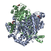
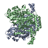
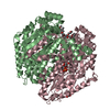
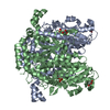
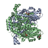
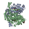
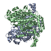
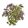
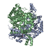
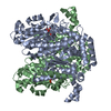
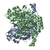
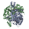
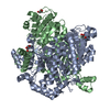
| Details | The biological unit is a dimer. The asymmetric unit is a dimer |
-Components
-Protein / Sugars , 2 types, 3 molecules AB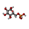
| #1: Protein | Mass: 63644.598 Da / Num. of mol.: 2 Source method: isolated from a genetically manipulated source Source: (gene. exp.)  #2: Sugar | ChemComp-G6P / |  Type: D-saccharide, alpha linking / Mass: 260.136 Da / Num. of mol.: 1 Type: D-saccharide, alpha linking / Mass: 260.136 Da / Num. of mol.: 1Source method: isolated from a genetically manipulated source Formula: C6H13O9P |
|---|
-Non-polymers , 5 types, 880 molecules 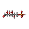


| #3: Chemical | ChemComp-G6Q /  Mass: 260.136 Da / Num. of mol.: 1 Mass: 260.136 Da / Num. of mol.: 1Source method: isolated from a genetically manipulated source Formula: C6H13O9P | ||||||
|---|---|---|---|---|---|---|---|
| #4: Chemical | ChemComp-SO4 /  Mass: 96.063 Da / Num. of mol.: 8 / Source method: obtained synthetically / Formula: SO4 Mass: 96.063 Da / Num. of mol.: 8 / Source method: obtained synthetically / Formula: SO4#5: Chemical | ChemComp-GOL /  Mass: 92.094 Da / Num. of mol.: 12 / Source method: obtained synthetically / Formula: C3H8O3 Mass: 92.094 Da / Num. of mol.: 12 / Source method: obtained synthetically / Formula: C3H8O3#6: Chemical | ChemComp-BME / |  Mass: 78.133 Da / Num. of mol.: 1 / Source method: obtained synthetically / Formula: C2H6OS Mass: 78.133 Da / Num. of mol.: 1 / Source method: obtained synthetically / Formula: C2H6OS#7: Water | ChemComp-HOH / | Mass: 18.015 Da / Num. of mol.: 858 / Source method: isolated from a natural source / Formula: H2O |
-Experimental details
-Experiment
| Experiment | Method: X-RAY DIFFRACTION / Number of used crystals: 1 |
|---|
- Sample preparation
| Crystal | Density Matthews: 2.3 Å3/Da / Density % sol: 41.5 % |
|---|---|
| Crystal grow | Temperature: 294 K / Method: vapor diffusion, hanging drop / pH: 8.5 Details: 1.9 M ammonium sulphate, 100 mM Tris-HCl, pH 8.5, VAPOR DIFFUSION, HANGING DROP, temperature 294K |
-Data collection
| Diffraction | Mean temperature: 100 K |
|---|---|
| Diffraction source | Source: SYNCHROTRON / Site: APS  / Beamline: 22-ID / Wavelength: 1 Å / Beamline: 22-ID / Wavelength: 1 Å |
| Detector | Type: MAR CCD 165 mm / Detector: CCD / Date: Aug 14, 2002 |
| Radiation | Protocol: SINGLE WAVELENGTH / Monochromatic (M) / Laue (L): M / Scattering type: x-ray |
| Radiation wavelength | Wavelength: 1 Å / Relative weight: 1 |
| Reflection | Resolution: 1.6→45 Å / Num. all: 143578 / Num. obs: 143578 / % possible obs: 94.7 % / Observed criterion σ(F): 0 / Observed criterion σ(I): 0 / Redundancy: 6 % / Biso Wilson estimate: 19.8 Å2 / Rmerge(I) obs: 0.057 / Rsym value: 0.057 / Net I/σ(I): 34.1 |
| Reflection shell | Resolution: 1.6→1.66 Å / Redundancy: 2.8 % / Rmerge(I) obs: 0.369 / Mean I/σ(I) obs: 3.1 / Num. unique all: 12096 / Rsym value: 0.369 / % possible all: 79.9 |
- Processing
| Software |
| ||||||||||||||||||||||||||||||||||||||||||||||||||||||||||||||||||||||||||||||||||||||||||||||||||||||||||||||||||||||||||||||||||||||||||||||||||||||||||||||||
|---|---|---|---|---|---|---|---|---|---|---|---|---|---|---|---|---|---|---|---|---|---|---|---|---|---|---|---|---|---|---|---|---|---|---|---|---|---|---|---|---|---|---|---|---|---|---|---|---|---|---|---|---|---|---|---|---|---|---|---|---|---|---|---|---|---|---|---|---|---|---|---|---|---|---|---|---|---|---|---|---|---|---|---|---|---|---|---|---|---|---|---|---|---|---|---|---|---|---|---|---|---|---|---|---|---|---|---|---|---|---|---|---|---|---|---|---|---|---|---|---|---|---|---|---|---|---|---|---|---|---|---|---|---|---|---|---|---|---|---|---|---|---|---|---|---|---|---|---|---|---|---|---|---|---|---|---|---|---|---|---|---|
| Refinement | Method to determine structure: REFINEMENT FROM THE NATIVE STRUCTURE Starting model: NATIVE STRUCTURE Resolution: 1.6→45 Å / Cor.coef. Fo:Fc: 0.967 / Cor.coef. Fo:Fc free: 0.956 / SU B: 1.787 / SU ML: 0.062 / Isotropic thermal model: ISOTROPIC / Cross valid method: THROUGHOUT / σ(F): 0 / ESU R: 0.095 / ESU R Free: 0.092 / Stereochemistry target values: MAXIMUM LIKELIHOOD
| ||||||||||||||||||||||||||||||||||||||||||||||||||||||||||||||||||||||||||||||||||||||||||||||||||||||||||||||||||||||||||||||||||||||||||||||||||||||||||||||||
| Solvent computation | Ion probe radii: 0.8 Å / Shrinkage radii: 0.8 Å / VDW probe radii: 1.4 Å / Solvent model: BABINET MODEL WITH MASK | ||||||||||||||||||||||||||||||||||||||||||||||||||||||||||||||||||||||||||||||||||||||||||||||||||||||||||||||||||||||||||||||||||||||||||||||||||||||||||||||||
| Displacement parameters | Biso mean: 23.854 Å2
| ||||||||||||||||||||||||||||||||||||||||||||||||||||||||||||||||||||||||||||||||||||||||||||||||||||||||||||||||||||||||||||||||||||||||||||||||||||||||||||||||
| Refinement step | Cycle: LAST / Resolution: 1.6→45 Å
| ||||||||||||||||||||||||||||||||||||||||||||||||||||||||||||||||||||||||||||||||||||||||||||||||||||||||||||||||||||||||||||||||||||||||||||||||||||||||||||||||
| Refine LS restraints |
| ||||||||||||||||||||||||||||||||||||||||||||||||||||||||||||||||||||||||||||||||||||||||||||||||||||||||||||||||||||||||||||||||||||||||||||||||||||||||||||||||
| LS refinement shell | Resolution: 1.6→1.642 Å / Total num. of bins used: 20
|
