+ Open data
- Basic information
| Entry | Database: PDB / ID: 1u0e | ||||||
|---|---|---|---|---|---|---|---|
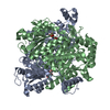
| Title | Crystal structure of mouse phosphoglucose isomerase | ||||||
 Components Components | Glucose-6-phosphate isomerase | ||||||
 Keywords Keywords | ISOMERASE / aldose-ketose isomerase / dimer | ||||||
| Function / homology |  Function and homology information Function and homology informationglycolytic process through glucose-6-phosphate / Gluconeogenesis / Glycolysis / TP53 Regulates Metabolic Genes / glucose-6-phosphate isomerase / glucose-6-phosphate isomerase activity / glucose 6-phosphate metabolic process / carbohydrate derivative binding / fructose 6-phosphate metabolic process / monosaccharide binding ...glycolytic process through glucose-6-phosphate / Gluconeogenesis / Glycolysis / TP53 Regulates Metabolic Genes / glucose-6-phosphate isomerase / glucose-6-phosphate isomerase activity / glucose 6-phosphate metabolic process / carbohydrate derivative binding / fructose 6-phosphate metabolic process / monosaccharide binding / canonical glycolysis / positive regulation of immunoglobulin production / ciliary membrane / erythrocyte homeostasis / response to testosterone / response to immobilization stress / mesoderm formation / response to cadmium ion / response to muscle stretch / Neutrophil degranulation / positive regulation of endothelial cell migration / response to progesterone / cytokine activity / glycolytic process / gluconeogenesis / growth factor activity / response to estradiol / myelin sheath / glucose homeostasis / in utero embryonic development / learning or memory / ubiquitin protein ligase binding / negative regulation of apoptotic process / extracellular space / plasma membrane / cytosol Similarity search - Function | ||||||
| Biological species |  | ||||||
| Method |  X-RAY DIFFRACTION / X-RAY DIFFRACTION /  MOLECULAR REPLACEMENT / Resolution: 1.6 Å MOLECULAR REPLACEMENT / Resolution: 1.6 Å | ||||||
 Authors Authors | Solomons, J.T.G. / Zimmerly, E.M. / Burns, S. / Krishnamurthy, N. / Swan, M.K. / Krings, S. / Muirhead, H. / Chirgwin, J. / Davies, C. | ||||||
 Citation Citation |  Journal: J.Mol.Biol. / Year: 2004 Journal: J.Mol.Biol. / Year: 2004Title: The crystal structure of mouse phosphoglucose isomerase at 1.6A resolution and its complex with glucose 6-phosphate reveals the catalytic mechanism of sugar ring opening. Authors: Graham Solomons, J.T. / Zimmerly, E.M. / Burns, S. / Krishnamurthy, N. / Swan, M.K. / Krings, S. / Muirhead, H. / Chirgwin, J. / Davies, C. | ||||||
| History |
| ||||||
| Remark 999 | SEQUENCE RESIDUE 263 IS A LEU NOT A PHE AS SHOWN BY THE SEQUENCE OF THE CONSTRUCT AS WELL AS THE ...SEQUENCE RESIDUE 263 IS A LEU NOT A PHE AS SHOWN BY THE SEQUENCE OF THE CONSTRUCT AS WELL AS THE ELECTRON DENSITY MAP. IT APPEARS TO EMANATE FROM THE ORIGINAL EXPRESSED SEQUENCE TAG AND IS NOT A PCR ERROR. HENCE THIS IS A POLYMORPHISM IN THE MAMMARY CELL LINE USED TO MAKE THE CDNA. |
- Structure visualization
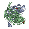
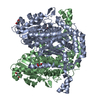
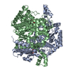
| Structure viewer | Molecule:  Molmil Molmil Jmol/JSmol Jmol/JSmol |
|---|
- Downloads & links
- Download
| PDBx/mmCIF format |  1u0e.cif.gz 1u0e.cif.gz | 252.8 KB | Display |  PDBx/mmCIF format PDBx/mmCIF format |
|---|---|---|---|---|
| PDB format |  pdb1u0e.ent.gz pdb1u0e.ent.gz | 200.6 KB | Display |  PDB format PDB format |
| PDBx/mmJSON format |  1u0e.json.gz 1u0e.json.gz | Tree view |  PDBx/mmJSON format PDBx/mmJSON format | |
| Others |  Other downloads Other downloads |
-Validation report
| Arichive directory |  https://data.pdbj.org/pub/pdb/validation_reports/u0/1u0e https://data.pdbj.org/pub/pdb/validation_reports/u0/1u0e ftp://data.pdbj.org/pub/pdb/validation_reports/u0/1u0e ftp://data.pdbj.org/pub/pdb/validation_reports/u0/1u0e | HTTPS FTP |
|---|
-Related structure data
| Related structure data | 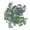 1u0fC  1u0gC  1n8tS C: citing same article ( S: Starting model for refinement |
|---|---|
| Similar structure data |
- Links
Links
- Assembly
| Deposited unit | 
| ||||||||
|---|---|---|---|---|---|---|---|---|---|
| 1 |
| ||||||||
| Unit cell |
| ||||||||
| Details | The biological unit is a dimer. The asymmetric unit is a dimer |
- Components
| #1: Protein | Mass: 63644.598 Da / Num. of mol.: 2 Source method: isolated from a genetically manipulated source Source: (gene. exp.)   #2: Chemical | ChemComp-SO4 / #3: Chemical | ChemComp-BME / #4: Chemical | ChemComp-GOL / #5: Water | ChemComp-HOH / | |
|---|
-Experimental details
-Experiment
| Experiment | Method:  X-RAY DIFFRACTION / Number of used crystals: 1 X-RAY DIFFRACTION / Number of used crystals: 1 |
|---|
- Sample preparation
| Crystal | Density Matthews: 2.3 Å3/Da / Density % sol: 41.5 % |
|---|---|
| Crystal grow | Temperature: 294 K / Method: vapor diffusion, hanging drop / pH: 8.5 Details: 1.9 M ammonium sulphate, 100 mM Tris-HCl, pH 8.5 , VAPOR DIFFUSION, HANGING DROP, temperature 294K |
-Data collection
| Diffraction | Mean temperature: 100 K |
|---|---|
| Diffraction source | Source:  ROTATING ANODE / Type: RIGAKU RU300 / Wavelength: 1.54 Å ROTATING ANODE / Type: RIGAKU RU300 / Wavelength: 1.54 Å |
| Detector | Type: RIGAKU RAXIS IV / Detector: IMAGE PLATE / Date: Jul 1, 2001 / Details: mirrors |
| Radiation | Monochromator: Osmic mirros / Protocol: SINGLE WAVELENGTH / Monochromatic (M) / Laue (L): M / Scattering type: x-ray |
| Radiation wavelength | Wavelength: 1.54 Å / Relative weight: 1 |
| Reflection | Resolution: 1.6→49.7 Å / Num. all: 148767 / Num. obs: 147857 / % possible obs: 99.5 % / Observed criterion σ(F): 0 / Observed criterion σ(I): 0 / Redundancy: 5.4 % / Biso Wilson estimate: 24.4 Å2 / Rmerge(I) obs: 0.075 / Rsym value: 0.075 / Net I/σ(I): 5.4 |
| Reflection shell | Resolution: 1.6→1.66 Å / Redundancy: 5.9 % / Rmerge(I) obs: 0.422 / Mean I/σ(I) obs: 1.5 / Num. unique all: 14817 / Rsym value: 0.422 / % possible all: 96.4 |
- Processing
| Software |
| ||||||||||||||||||||||||||||||||||||||||||||||||||||||||||||||||||||||||||||||||||||||||||||||||||||||||||||||||||||||||||||||||||||||||||||||||||||||||||||||||
|---|---|---|---|---|---|---|---|---|---|---|---|---|---|---|---|---|---|---|---|---|---|---|---|---|---|---|---|---|---|---|---|---|---|---|---|---|---|---|---|---|---|---|---|---|---|---|---|---|---|---|---|---|---|---|---|---|---|---|---|---|---|---|---|---|---|---|---|---|---|---|---|---|---|---|---|---|---|---|---|---|---|---|---|---|---|---|---|---|---|---|---|---|---|---|---|---|---|---|---|---|---|---|---|---|---|---|---|---|---|---|---|---|---|---|---|---|---|---|---|---|---|---|---|---|---|---|---|---|---|---|---|---|---|---|---|---|---|---|---|---|---|---|---|---|---|---|---|---|---|---|---|---|---|---|---|---|---|---|---|---|---|
| Refinement | Method to determine structure:  MOLECULAR REPLACEMENT MOLECULAR REPLACEMENTStarting model: PDB ENTRY 1N8T Resolution: 1.6→14.96 Å / Cor.coef. Fo:Fc: 0.962 / Cor.coef. Fo:Fc free: 0.946 / SU B: 2.67 / SU ML: 0.086 / Isotropic thermal model: isotropic / Cross valid method: THROUGHOUT / σ(F): 0 / ESU R: 0.109 / ESU R Free: 0.108 / Stereochemistry target values: MAXIMUM LIKELIHOOD
| ||||||||||||||||||||||||||||||||||||||||||||||||||||||||||||||||||||||||||||||||||||||||||||||||||||||||||||||||||||||||||||||||||||||||||||||||||||||||||||||||
| Solvent computation | Ion probe radii: 0.8 Å / Shrinkage radii: 0.8 Å / VDW probe radii: 1.4 Å / Solvent model: BABINET MODEL WITH MASK | ||||||||||||||||||||||||||||||||||||||||||||||||||||||||||||||||||||||||||||||||||||||||||||||||||||||||||||||||||||||||||||||||||||||||||||||||||||||||||||||||
| Displacement parameters | Biso mean: 24.439 Å2
| ||||||||||||||||||||||||||||||||||||||||||||||||||||||||||||||||||||||||||||||||||||||||||||||||||||||||||||||||||||||||||||||||||||||||||||||||||||||||||||||||
| Refinement step | Cycle: LAST / Resolution: 1.6→14.96 Å
| ||||||||||||||||||||||||||||||||||||||||||||||||||||||||||||||||||||||||||||||||||||||||||||||||||||||||||||||||||||||||||||||||||||||||||||||||||||||||||||||||
| Refine LS restraints |
| ||||||||||||||||||||||||||||||||||||||||||||||||||||||||||||||||||||||||||||||||||||||||||||||||||||||||||||||||||||||||||||||||||||||||||||||||||||||||||||||||
| LS refinement shell | Resolution: 1.6→1.641 Å / Total num. of bins used: 20
|
 Movie
Movie Controller





 PDBj
PDBj


