 Movie
Movie Controller

[English] 日本語
 Yorodumi
- PDB-1iri: Crystal structure of human autocrine motility factor complexed wi... -
+ Open data

- Basic information
| Entry | Database: PDB / ID: 1iri | ||||||
|---|---|---|---|---|---|---|---|
| Title | Crystal structure of human autocrine motility factor complexed with an inhibitor | ||||||
 Components Components | autocrine motility factor | ||||||
 Keywords Keywords | ISOMERASE / cytokine | ||||||
| Function / homology |  Function and homology information Function and homology informationIsomerases; Racemases and epimerases; Acting on carbohydrates and derivatives / racemase and epimerase activity, acting on carbohydrates and derivatives / glucose-6-phosphate isomerase / glucose-6-phosphate isomerase activity / hemostasis / glucose 6-phosphate metabolic process / carbohydrate derivative binding / fructose 6-phosphate metabolic process / monosaccharide binding / Gluconeogenesis ...Isomerases; Racemases and epimerases; Acting on carbohydrates and derivatives / racemase and epimerase activity, acting on carbohydrates and derivatives / glucose-6-phosphate isomerase / glucose-6-phosphate isomerase activity / hemostasis / glucose 6-phosphate metabolic process / carbohydrate derivative binding / fructose 6-phosphate metabolic process / monosaccharide binding / Gluconeogenesis / Glycolysis / canonical glycolysis / ciliary membrane / positive regulation of immunoglobulin production / response to testosterone / erythrocyte homeostasis / negative regulation of glycolytic process through fructose-6-phosphate / response to immobilization stress / humoral immune response / mesoderm formation / response to cadmium ion / response to muscle stretch / response to progesterone / positive regulation of endothelial cell migration / cytokine activity / glycolytic process / gluconeogenesis / TP53 Regulates Metabolic Genes / growth factor activity / response to estradiol / glucose homeostasis / secretory granule lumen / carbohydrate metabolic process / ficolin-1-rich granule lumen / in utero embryonic development / learning or memory / Neutrophil degranulation / ubiquitin protein ligase binding / negative regulation of apoptotic process / extracellular exosome / extracellular region / membrane / cytosol Similarity search - Function | ||||||
| Biological species |  Homo sapiens (human) Homo sapiens (human) | ||||||
| Method |  X-RAY DIFFRACTION / SYNCHROTRON / FOURIER SYNTHESIS / Resolution: 2.4 Å X-RAY DIFFRACTION / SYNCHROTRON / FOURIER SYNTHESIS / Resolution: 2.4 Å | ||||||
 Authors Authors | Tanaka, N. / Haga, A. / Uemura, H. / Akiyama, H. / Funasaka, T. / Nagase, H. / Raz, A. / Nakamura, K.T. | ||||||
 Citation Citation | Journal: J.Mol.Biol. / Year: 2002 Title: Inhibition mechanism of cytokine activity of human autocrine motility factor examined by crystal structure analyses and site-directed mutagenesis studies. Authors: Tanaka, N. / Haga, A. / Uemura, H. / Akiyama, H. / Funasaka, T. / Nagase, H. / Raz, A. / Nakamura, K.T. | ||||||
| History |
|
- Structure visualization
| Structure viewer | Molecule: MolmilJmol/JSmol |
|---|
- Downloads & links
-Download
| PDBx/mmCIF format | 1iri.cif.gz | 440.9 KB | Display | PDBx/mmCIF format |
|---|---|---|---|---|
| PDB format | pdb1iri.ent.gz | 365.1 KB | Display | PDB format |
| PDBx/mmJSON format | 1iri.json.gz | Tree view | PDBx/mmJSON format | |
| Others |  Other downloads Other downloads |
-Validation report
| Arichive directory | https://data.pdbj.org/pub/pdb/validation_reports/ir/1iriftp://data.pdbj.org/pub/pdb/validation_reports/ir/1iri | HTTPS FTP |
|---|
-Related structure data
| Related structure data |  1jiqSC S: Starting model for refinement C: citing same article ( |
|---|---|
| Similar structure data |


-Links
 PDBj
PDBj


- Assembly
| Deposited unit | 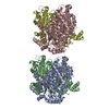
| ||||||||
|---|---|---|---|---|---|---|---|---|---|
| 1 | 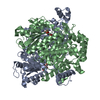
| ||||||||
| 2 | 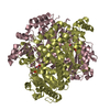
| ||||||||
| Unit cell |
| ||||||||
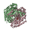
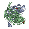
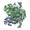
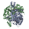
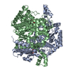
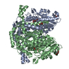
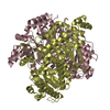
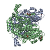
| Details | The biological assembly is a dimer. Two dimers exist in an asymmetric unit. |
-Components
| #1: Protein | Mass: 63229.949 Da / Num. of mol.: 4 Source method: isolated from a genetically manipulated source Source: (gene. exp.) Homo sapiens (human) / Plasmid: pGEX-6P / Production host:  #2: Sugar | ChemComp-E4P / 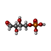  Type: saccharide / Mass: 200.084 Da / Num. of mol.: 4 / Source method: obtained synthetically / Formula: C4H9O7P Type: saccharide / Mass: 200.084 Da / Num. of mol.: 4 / Source method: obtained synthetically / Formula: C4H9O7P#3: Water | ChemComp-HOH / |  Mass: 18.015 Da / Num. of mol.: 432 / Source method: isolated from a natural source / Formula: H2O Mass: 18.015 Da / Num. of mol.: 432 / Source method: isolated from a natural source / Formula: H2O |
|---|
-Experimental details
-Experiment
| Experiment | Method: X-RAY DIFFRACTION / Number of used crystals: 1 |
|---|
- Sample preparation
| Crystal | Density Matthews: 2.32 Å3/Da / Density % sol: 47.02 % | |||||||||||||||||||||||||||||||||||||||||||||||||||||||||||||||
|---|---|---|---|---|---|---|---|---|---|---|---|---|---|---|---|---|---|---|---|---|---|---|---|---|---|---|---|---|---|---|---|---|---|---|---|---|---|---|---|---|---|---|---|---|---|---|---|---|---|---|---|---|---|---|---|---|---|---|---|---|---|---|---|---|
| Crystal grow | Temperature: 293 K / Method: vapor diffusion, hanging drop / pH: 6.5 Details: cacodylate, sodium acetate, PEG8000, pH 6.5, VAPOR DIFFUSION, HANGING DROP, temperature 293K | |||||||||||||||||||||||||||||||||||||||||||||||||||||||||||||||
| Crystal | *PLUS Density % sol: 47 % | |||||||||||||||||||||||||||||||||||||||||||||||||||||||||||||||
| Crystal grow | *PLUS pH: 7.5 | |||||||||||||||||||||||||||||||||||||||||||||||||||||||||||||||
| Components of the solutions | *PLUS
|
-Data collection
| Diffraction | Mean temperature: 100 K |
|---|---|
| Diffraction source | Source: SYNCHROTRON / Site: Photon Factory  / Beamline: BL-6A / Wavelength: 1 Å / Beamline: BL-6A / Wavelength: 1 Å |
| Detector | Type: ADSC QUANTUM 4 / Detector: CCD |
| Radiation | Monochromator: Si 111 / Protocol: SINGLE WAVELENGTH / Monochromatic (M) / Laue (L): M / Scattering type: x-ray |
| Radiation wavelength | Wavelength: 1 Å / Relative weight: 1 |
| Reflection | Resolution: 2.4→25 Å / Num. all: 92870 / Num. obs: 92870 / % possible obs: 100 % / Redundancy: 5.9 % / Rmerge(I) obs: 0.09 / Net I/σ(I): 7.3 |
| Reflection shell | Resolution: 2→2.53 Å / Redundancy: 6 % / Rmerge(I) obs: 0.357 / Mean I/σ(I) obs: 2.1 / % possible all: 99.9 |
| Reflection | *PLUS Lowest resolution: 25 Å / % possible obs: 100 % / Num. measured all: 550782 / Rmerge(I) obs: 0.09 |
| Reflection shell | *PLUS Highest resolution: 2.4 Å / % possible obs: 99.9 % / Rmerge(I) obs: 0.357 |
- Processing
| Software |
| ||||||||||||||||
|---|---|---|---|---|---|---|---|---|---|---|---|---|---|---|---|---|---|
| Refinement | Method to determine structure: FOURIER SYNTHESIS Starting model: PDB ENTRY 1JIQ Resolution: 2.4→25 Å
| ||||||||||||||||
| Refinement step | Cycle: LAST / Resolution: 2.4→25 Å
| ||||||||||||||||
| Refine LS restraints | Type: p_bond_d / Dev ideal: 0.013 | ||||||||||||||||
| Refinement | *PLUS Lowest resolution: 25 Å / Rfactor obs: 0.193 / Rfactor Rfree: 0.241 / Rfactor Rwork: 0.193 | ||||||||||||||||
| Solvent computation | *PLUS | ||||||||||||||||
| Displacement parameters | *PLUS | ||||||||||||||||
| Refine LS restraints | *PLUS
| ||||||||||||||||
| LS refinement shell | *PLUS Highest resolution: 2.4 Å / Lowest resolution: 2.46 Å |
