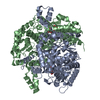
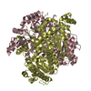
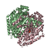
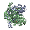
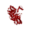
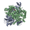
[English] 日本語
Yorodumi
- PDB-4wmj: Colias eurytheme Phosphoglucose isomerase. Homodimer from 4-5(18)... -

+
Open data


ID or keywords:

Loading...

-
Basic information

Entry
Database: PDB / ID: 4wmj
TitleColias eurytheme Phosphoglucose isomerase. Homodimer from 4-5(18) genotype.
ComponentsGlucose-6-phosphate isomerase
KeywordsISOMERASE / Glycolysis
Function / homology
Function and homology information


glucose-6-phosphate isomerase / glucose-6-phosphate isomerase activity / gluconeogenesis / glycolytic process
Similarity search - Function
Phosphoglucose isomerase, C-terminal domain / Phosphoglucose isomerase, C-terminal domain - #10 / Phosphoglucose isomerase, C-terminal / Phosphoglucose isomerase signature 1. / Phosphoglucose isomerase (PGI) / Phosphoglucose isomerase, conserved site / Phosphoglucose isomerase, SIS domain 1 / Phosphoglucose isomerase, SIS domain 2 / Phosphoglucose isomerase / Phosphoglucose isomerase signature 2. ...Phosphoglucose isomerase, C-terminal domain / Phosphoglucose isomerase, C-terminal domain - #10 / Phosphoglucose isomerase, C-terminal / Phosphoglucose isomerase signature 1. / Phosphoglucose isomerase (PGI) / Phosphoglucose isomerase, conserved site / Phosphoglucose isomerase, SIS domain 1 / Phosphoglucose isomerase, SIS domain 2 / Phosphoglucose isomerase / Phosphoglucose isomerase signature 2. / Glucose-6-phosphate isomerase family profile. / Glucose-6-phosphate isomerase like protein; domain 1 / Rossmann fold / Orthogonal Bundle / 3-Layer(aba) Sandwich / Mainly Alpha / Alpha Beta
Similarity search - Domain/homology
Glucose-6-phosphate isomerase
Similarity search - Component
Biological speciesColias eurytheme (orange sulphur)
MethodX-RAY DIFFRACTION / SYNCHROTRON / MOLECULAR REPLACEMENT / Resolution: 1.5 Å
AuthorsHill, J.A. / Watt, W.B.
CitationJournal: Thesis, Stanford University / Year: 2013
Title: Structure and Function of Phosphoglucose Isomerase In Colias Eurytheme
Authors: Hill, J.A. / Watt, W.B. / Jardetzky, T. / Tuljapurkar, S.
History
DepositionOct 9, 2014Deposition site: RCSB / Processing site: RCSB
Revision 1.0Oct 14, 2015Provider: repository / Type: Initial release
Revision 1.1Sep 27, 2023Group: Data collection / Database references ...Data collection / Database references / Derived calculations / Refinement description
Category: chem_comp_atom / chem_comp_bond ...chem_comp_atom / chem_comp_bond / database_2 / pdbx_initial_refinement_model / pdbx_struct_oper_list
Item: _database_2.pdbx_DOI / _database_2.pdbx_database_accession / _pdbx_struct_oper_list.symmetry_operation

-
Structure visualization

Structure viewerMolecule:
MolmilJmol/JSmol

Downloads & links

-
Assembly

Deposited unit
A: Glucose-6-phosphate isomerase
B: Glucose-6-phosphate isomerase
C: Glucose-6-phosphate isomerase
D: Glucose-6-phosphate isomerase


Theoretical massNumber of molelcules
Total (without water)247,7534
Polymers247,7534
Non-polymers00
Water19,6901093
1
A: Glucose-6-phosphate isomerase
B: Glucose-6-phosphate isomerase


Theoretical massNumber of molelcules
Total (without water)123,8762
Polymers123,8762
Non-polymers00
Water362
TypeNameSymmetry operationNumber
identity operation1_555x,y,z1
Buried area12410 Å2
ΔGint-66 kcal/mol
Surface area36220 Å2
MethodPISA
2
C: Glucose-6-phosphate isomerase
D: Glucose-6-phosphate isomerase


Theoretical massNumber of molelcules
Total (without water)123,8762
Polymers123,8762
Non-polymers00
Water362
TypeNameSymmetry operationNumber
identity operation1_555x,y,z1
Buried area12390 Å2
ΔGint-67 kcal/mol
Surface area36140 Å2
MethodPISA
Unit cell
Length a, b, c (Å)76.287, 115.495, 135.079
Angle α, β, γ (deg.)90.00, 101.70, 90.00
Int Tables number4
Space group name H-MP1211

-
Components

#1: Protein
Glucose-6-phosphate isomerase /


Mass: 61938.219 Da / Num. of mol.: 4
Source method: isolated from a genetically manipulated source
Source: (gene. exp.) Colias eurytheme (orange sulphur) / Gene: PGI / Production host: Escherichia coli (E. coli) / References: UniProt: G9BZJ0, glucose-6-phosphate isomerase
#2: Water ChemComp-HOH / water / Water


Mass: 18.015 Da / Num. of mol.: 1093 / Source method: isolated from a natural source / Formula: H2O

-
Experimental details

-
Experiment

ExperimentMethod: X-RAY DIFFRACTION / Number of used crystals: 1

-
Sample preparation

CrystalDensity Matthews: 2.35 Å3/Da / Density % sol: 47.7 %
Crystal growTemperature: 297 K / Method: vapor diffusion, hanging drop / pH: 7.4
Details: 0.1M lithium sulfate, 22% PEG 4000, 0.2M Tris-Cl pH 7.4.

-
Data collection

DiffractionMean temperature: 80 K
Diffraction sourceSource: SYNCHROTRON / Site: SSRL / Beamline: BL11-1 / Wavelength: 0.98 Å
DetectorType: DECTRIS PILATUS 6M / Detector: PIXEL / Date: Jul 15, 2011
RadiationProtocol: SINGLE WAVELENGTH / Monochromatic (M) / Laue (L): M / Scattering type: x-ray
Radiation wavelengthWavelength: 0.98 Å / Relative weight: 1
ReflectionResolution: 1.5→41.19 Å / Num. obs: 270174 / % possible obs: 77.68 % / Redundancy: 2 % / Net I/σ(I): 8.99

-
Processing

Software
NameVersionClassification
REFMAC5.8.0073refinement
MOLREPphasing
HKL-3000data collection
HKL-3000data reduction
HKL-3000data scaling
RefinementMethod to determine structure: MOLECULAR REPLACEMENT
Starting model: 1U0E
Resolution: 1.5→41.19 Å / Cor.coef. Fo:Fc: 0.96 / Cor.coef. Fo:Fc free: 0.943 / SU B: 4.507 / SU ML: 0.078 / Cross valid method: THROUGHOUT / ESU R: 0.093 / ESU R Free: 0.095 / Stereochemistry target values: MAXIMUM LIKELIHOOD / Details: HYDROGENS HAVE BEEN ADDED IN THE RIDING POSITIONS
RfactorNum. reflection% reflectionSelection details
Rfree0.2179 14357 5 %RANDOM
Rwork0.18315 ---
obs0.1849 270174 77.68 %-
Solvent computationIon probe radii: 0.8 Å / Shrinkage radii: 0.8 Å / VDW probe radii: 1.2 Å / Solvent model: MASK
Displacement parametersBiso mean: 18.161 Å2
Baniso -1Baniso -2Baniso -3
1--0.13 Å20 Å2-0.01 Å2
2--0.23 Å20 Å2
3----0.09 Å2
Refinement stepCycle: LAST / Resolution: 1.5→41.19 Å
ProteinNucleic acidLigandSolventTotal
Num. atoms17415 0 0 1093 18508
Refine LS restraints
Refine-IDTypeDev idealDev ideal targetNumber
X-RAY DIFFRACTIONr_bond_refined_d0.020.01917845
X-RAY DIFFRACTIONr_bond_other_d0.0010.0216984
X-RAY DIFFRACTIONr_angle_refined_deg1.91.94224215
X-RAY DIFFRACTIONr_angle_other_deg0.93339058
X-RAY DIFFRACTIONr_dihedral_angle_1_deg6.35952211
X-RAY DIFFRACTIONr_dihedral_angle_2_deg38.28524.909825
X-RAY DIFFRACTIONr_dihedral_angle_3_deg13.867152974
X-RAY DIFFRACTIONr_dihedral_angle_4_deg18.6541560
X-RAY DIFFRACTIONr_chiral_restr0.1240.22663
X-RAY DIFFRACTIONr_gen_planes_refined0.0110.02120410
X-RAY DIFFRACTIONr_gen_planes_other0.0010.024214
X-RAY DIFFRACTIONr_nbd_refined
X-RAY DIFFRACTIONr_nbd_other
X-RAY DIFFRACTIONr_nbtor_refined
X-RAY DIFFRACTIONr_nbtor_other
X-RAY DIFFRACTIONr_xyhbond_nbd_refined
X-RAY DIFFRACTIONr_xyhbond_nbd_other
X-RAY DIFFRACTIONr_metal_ion_refined
X-RAY DIFFRACTIONr_metal_ion_other
X-RAY DIFFRACTIONr_symmetry_vdw_refined
X-RAY DIFFRACTIONr_symmetry_vdw_other
X-RAY DIFFRACTIONr_symmetry_hbond_refined
X-RAY DIFFRACTIONr_symmetry_hbond_other
X-RAY DIFFRACTIONr_symmetry_metal_ion_refined
X-RAY DIFFRACTIONr_symmetry_metal_ion_other
X-RAY DIFFRACTIONr_mcbond_it1.3081.3898856
X-RAY DIFFRACTIONr_mcbond_other1.3081.3898855
X-RAY DIFFRACTIONr_mcangle_it1.8532.0811063
X-RAY DIFFRACTIONr_mcangle_other1.8532.0811064
X-RAY DIFFRACTIONr_scbond_it2.4411.6228989
X-RAY DIFFRACTIONr_scbond_other2.4411.6228990
X-RAY DIFFRACTIONr_scangle_it
X-RAY DIFFRACTIONr_scangle_other3.6232.32613153
X-RAY DIFFRACTIONr_long_range_B_refined4.4211.76621716
X-RAY DIFFRACTIONr_long_range_B_other4.39911.60621275
X-RAY DIFFRACTIONr_rigid_bond_restr
X-RAY DIFFRACTIONr_sphericity_free
X-RAY DIFFRACTIONr_sphericity_bonded
LS refinement shellResolution: 1.5→1.537 Å / Total num. of bins used: 20
RfactorNum. reflection% reflection
Rfree0.423 294 -
Rwork0.4 5376 -
obs--21.02 %
Refinement TLS params.

Method: refined / Refine-ID: X-RAY DIFFRACTION

IDL112)L122)L132)L222)L232)L332)S11 (Å °)S12 (Å °)S13 (Å °)S21 (Å °)S22 (Å °)S23 (Å °)S31 (Å °)S32 (Å °)S33 (Å °)T112)T122)T132)T222)T232)T332)Origin x (Å)Origin y (Å)Origin z (Å)
10.08730.03730.07730.06710.0340.16680.02430.0143-0.0026-0.007-0.013-0.00110.01580.0307-0.01140.01650.0072-0.00250.03190.00030.0024-33.7174-2.028722.7841
20.0637-0.00650.04360.13420.00890.1560.0205-0.00670.00050.0093-0.01360.03070.0002-0.0373-0.0070.0095-0.00310.00420.0387-0.00170.008-54.52241.499334.3117
30.05760.0372-0.01080.1674-0.03680.0962-0.0142-0.00740.0073-0.01380.02920.00750.039-0.0261-0.0150.0181-0.0052-0.0070.03480.00250.0028-28.5711-10.770696.8076
40.04790.0044-0.00110.1116-0.0530.1479-0.0076-0.00610.0175-0.0050.0099-0.0286-0.0092-0.018-0.00230.00820.0072-0.00030.0289-0.00160.017-17.13669.136789.6017
Refinement TLS group
IDRefine-IDRefine TLS-IDAuth asym-IDAuth seq-ID
1X-RAY DIFFRACTION1A4 - 556
2X-RAY DIFFRACTION2B2 - 556
3X-RAY DIFFRACTION3C3 - 556
4X-RAY DIFFRACTION4D4 - 556

+
About Yorodumi

-
News

-
Feb 9, 2022. New format data for meta-information of EMDB entries

New format data for meta-information of EMDB entries

  • Version 3 of the EMDB header file is now the official format.
  • The previous official version 1.9 will be removed from the archive.

Related info.:EMDB header

External links:wwPDB to switch to version 3 of the EMDB data model

-
Aug 12, 2020. Covid-19 info

Covid-19 info

URL: https://pdbj.org/emnavi/covid19.php

New page: Covid-19 featured information page in EM Navigator.

Related info.:Covid-19 info / Mar 5, 2020. Novel coronavirus structure data

+
Mar 5, 2020. Novel coronavirus structure data

Novel coronavirus structure data

Related info.:Yorodumi Speices / Aug 12, 2020. Covid-19 info

External links:COVID-19 featured content - PDBj / Molecule of the Month (242):Coronavirus Proteases

+
Jan 31, 2019. EMDB accession codes are about to change! (news from PDBe EMDB page)

EMDB accession codes are about to change! (news from PDBe EMDB page)

  • The allocation of 4 digits for EMDB accession codes will soon come to an end. Whilst these codes will remain in use, new EMDB accession codes will include an additional digit and will expand incrementally as the available range of codes is exhausted. The current 4-digit format prefixed with “EMD-” (i.e. EMD-XXXX) will advance to a 5-digit format (i.e. EMD-XXXXX), and so on. It is currently estimated that the 4-digit codes will be depleted around Spring 2019, at which point the 5-digit format will come into force.
  • The EM Navigator/Yorodumi systems omit the EMD- prefix.

Related info.:Q: What is EMD? / ID/Accession-code notation in Yorodumi/EM Navigator

External links:EMDB Accession Codes are Changing Soon! / Contact to PDBj

+
Jul 12, 2017. Major update of PDB

Major update of PDB

  • wwPDB released updated PDB data conforming to the new PDBx/mmCIF dictionary.
  • This is a major update changing the version number from 4 to 5, and with Remediation, in which all the entries are updated.
  • In this update, many items about electron microscopy experimental information are reorganized (e.g. em_software).
  • Now, EM Navigator and Yorodumi are based on the updated data.

External links:wwPDB Remediation / Enriched Model Files Conforming to OneDep Data Standards Now Available in the PDB FTP Archive

-
Yorodumi

Thousand views of thousand structures

  • Yorodumi is a browser for structure data from EMDB, PDB, SASBDB, etc.
  • This page is also the successor to EM Navigator detail page, and also detail information page/front-end page for Omokage search.
  • The word "yorodu" (or yorozu) is an old Japanese word meaning "ten thousand". "mi" (miru) is to see.

Related info.:EMDB / PDB / SASBDB / Comparison of 3 databanks / Yorodumi Search / Aug 31, 2016. New EM Navigator & Yorodumi / Yorodumi Papers / Jmol/JSmol / Function and homology information / Changes in new EM Navigator and Yorodumi

Read more