 Movie
Movie Controller

[English] 日本語
 Yorodumi
- PDB-1iat: CRYSTAL STRUCTURE OF HUMAN PHOSPHOGLUCOSE ISOMERASE/NEUROLEUKIN/A... -
+ Open data

- Basic information
| Entry | Database: PDB / ID: 1iat | ||||||
|---|---|---|---|---|---|---|---|
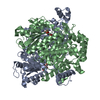
| Title | CRYSTAL STRUCTURE OF HUMAN PHOSPHOGLUCOSE ISOMERASE/NEUROLEUKIN/AUTOCRINE MOTILITY FACTOR/MATURATION FACTOR | ||||||
 Components Components | PHOSPHOGLUCOSE ISOMERASE | ||||||
 Keywords Keywords | ISOMERASE / glycolysis enzyme/neurotrophic growth factor/cytokine / two alpha/beta domains | ||||||
| Function / homology |  Function and homology information Function and homology informationIsomerases; Racemases and epimerases; Acting on carbohydrates and derivatives / racemase and epimerase activity, acting on carbohydrates and derivatives / glucose-6-phosphate isomerase / glucose-6-phosphate isomerase activity / hemostasis / glucose 6-phosphate metabolic process / carbohydrate derivative binding / fructose 6-phosphate metabolic process / monosaccharide binding / Gluconeogenesis ...Isomerases; Racemases and epimerases; Acting on carbohydrates and derivatives / racemase and epimerase activity, acting on carbohydrates and derivatives / glucose-6-phosphate isomerase / glucose-6-phosphate isomerase activity / hemostasis / glucose 6-phosphate metabolic process / carbohydrate derivative binding / fructose 6-phosphate metabolic process / monosaccharide binding / Gluconeogenesis / Glycolysis / canonical glycolysis / ciliary membrane / response to testosterone / erythrocyte homeostasis / positive regulation of immunoglobulin production / negative regulation of glycolytic process through fructose-6-phosphate / response to immobilization stress / humoral immune response / mesoderm formation / response to cadmium ion / response to muscle stretch / response to progesterone / positive regulation of endothelial cell migration / cytokine activity / gluconeogenesis / glycolytic process / TP53 Regulates Metabolic Genes / growth factor activity / response to estradiol / glucose homeostasis / in utero embryonic development / secretory granule lumen / carbohydrate metabolic process / ficolin-1-rich granule lumen / learning or memory / ubiquitin protein ligase binding / Neutrophil degranulation / negative regulation of apoptotic process / extracellular exosome / extracellular region / membrane / cytosol Similarity search - Function | ||||||
| Biological species |  Homo sapiens (human) Homo sapiens (human) | ||||||
| Method |  X-RAY DIFFRACTION / SYNCHROTRON / MOLECULAR REPLACEMENT / Resolution: 1.62 Å X-RAY DIFFRACTION / SYNCHROTRON / MOLECULAR REPLACEMENT / Resolution: 1.62 Å | ||||||
 Authors Authors | Read, J.A. / Pearce, J. / Li, X. / Muirhead, H. / Chirgwin, J. / Davies, C. | ||||||
 Citation Citation | Journal: J.Mol.Biol. / Year: 2001 Title: The crystal structure of human phosphoglucose isomerase at 1.6 A resolution: implications for catalytic mechanism, cytokine activity and haemolytic anaemia. Authors: Read, J. / Pearce, J. / Li, X. / Muirhead, H. / Chirgwin, J. / Davies, C. | ||||||
| History |
|
- Structure visualization
| Structure viewer | Molecule: MolmilJmol/JSmol |
|---|
- Downloads & links
-Download
| PDBx/mmCIF format | 1iat.cif.gz | 247.1 KB | Display | PDBx/mmCIF format |
|---|---|---|---|---|
| PDB format | pdb1iat.ent.gz | 199.2 KB | Display | PDB format |
| PDBx/mmJSON format | 1iat.json.gz | Tree view | PDBx/mmJSON format | |
| Others |  Other downloads Other downloads |
-Validation report
| Arichive directory | https://data.pdbj.org/pub/pdb/validation_reports/ia/1iatftp://data.pdbj.org/pub/pdb/validation_reports/ia/1iat | HTTPS FTP |
|---|
-Related structure data
| Related structure data | |
|---|---|
| Similar structure data |


-Links
 PDBj
PDBj


- Assembly
| Deposited unit | 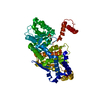
| |||||||||
|---|---|---|---|---|---|---|---|---|---|---|
| 1 | 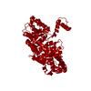
| |||||||||
| Unit cell |
| |||||||||
| Components on special symmetry positions |
|
-Components
| #1: Protein | Mass: 63098.746 Da / Num. of mol.: 1 Source method: isolated from a genetically manipulated source Source: (gene. exp.) Homo sapiens (human) / Gene: GPI / Production host:  | ||||
|---|---|---|---|---|---|
| #2: Chemical |   Mass: 96.063 Da / Num. of mol.: 2 / Source method: obtained synthetically / Formula: SO4 Mass: 96.063 Da / Num. of mol.: 2 / Source method: obtained synthetically / Formula: SO4#3: Chemical | ChemComp-BME / |   Mass: 78.133 Da / Num. of mol.: 1 / Source method: obtained synthetically / Formula: C2H6OS Mass: 78.133 Da / Num. of mol.: 1 / Source method: obtained synthetically / Formula: C2H6OS#4: Water | ChemComp-HOH / |  Mass: 18.015 Da / Num. of mol.: 581 / Source method: isolated from a natural source / Formula: H2O Mass: 18.015 Da / Num. of mol.: 581 / Source method: isolated from a natural source / Formula: H2O |
-Experimental details
-Experiment
| Experiment | Method: X-RAY DIFFRACTION / Number of used crystals: 1 |
|---|
- Sample preparation
| Crystal | Density Matthews: 2.39 Å3/Da / Density % sol: 48.57 % | ||||||||||||||||||||||||||||||
|---|---|---|---|---|---|---|---|---|---|---|---|---|---|---|---|---|---|---|---|---|---|---|---|---|---|---|---|---|---|---|---|
| Crystal grow | Temperature: 291 K / Method: vapor diffusion, hanging drop / pH: 8.5 Details: Ammonium sulphate, tris-HCl, Na Hepes, beta-mercaptoethanol, pH 8.5, VAPOR DIFFUSION, HANGING DROP, temperature 291K | ||||||||||||||||||||||||||||||
| Crystal grow | *PLUS pH: 7.5 | ||||||||||||||||||||||||||||||
| Components of the solutions | *PLUS
|
-Data collection
| Diffraction | Mean temperature: 100 K |
|---|---|
| Diffraction source | Source: SYNCHROTRON / Site: SRS  / Beamline: PX7.2 / Wavelength: 1.488 Å / Beamline: PX7.2 / Wavelength: 1.488 Å |
| Detector | Type: MARRESEARCH / Detector: IMAGE PLATE / Date: Mar 17, 2000 |
| Radiation | Protocol: SINGLE WAVELENGTH / Monochromatic (M) / Laue (L): M / Scattering type: x-ray |
| Radiation wavelength | Wavelength: 1.488 Å / Relative weight: 1 |
| Reflection | Resolution: 1.62→30 Å / Num. all: 75504 / Num. obs: 75504 / % possible obs: 96.4 % / Observed criterion σ(F): 0 / Observed criterion σ(I): 0 / Redundancy: 5.13 % / Biso Wilson estimate: 14.3 Å2 / Rmerge(I) obs: 0.048 / Net I/σ(I): 30.65 |
| Reflection shell | Resolution: 1.62→1.69 Å / Redundancy: 4.52 % / Rmerge(I) obs: 0.173 / Mean I/σ(I) obs: 7.33 / % possible all: 88.4 |
| Reflection | *PLUS |
| Reflection shell | *PLUS % possible obs: 88.4 % |
- Processing
| Software |
| |||||||||||||||||||||||||
|---|---|---|---|---|---|---|---|---|---|---|---|---|---|---|---|---|---|---|---|---|---|---|---|---|---|---|
| Refinement | Method to determine structure: MOLECULAR REPLACEMENT Starting model: PIG MUSCLE PGI (unpublished) Resolution: 1.62→29.88 Å / Cross valid method: THROUGHOUT / σ(F): 0 / σ(I): 0 / Stereochemistry target values: Engh & Huber
| |||||||||||||||||||||||||
| Displacement parameters | Biso mean: 17.1 Å2
| |||||||||||||||||||||||||
| Refinement step | Cycle: LAST / Resolution: 1.62→29.88 Å
| |||||||||||||||||||||||||
| Refine LS restraints |
| |||||||||||||||||||||||||
| LS refinement shell | Resolution: 1.62→1.662 Å / Total num. of bins used: 20 /
| |||||||||||||||||||||||||
| Software | *PLUS Name: REFMAC / Version: 5 / Classification: refinement | |||||||||||||||||||||||||
| Refinement | *PLUS σ(F): 0 / % reflection Rfree: 5 % / Rfactor Rfree: 0.17 | |||||||||||||||||||||||||
| Solvent computation | *PLUS | |||||||||||||||||||||||||
| Displacement parameters | *PLUS Biso mean: 17.1 Å2 | |||||||||||||||||||||||||
| Refine LS restraints | *PLUS
|
