 Movie
Movie Controller

[English] 日本語
 Yorodumi
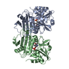
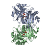
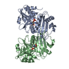
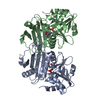
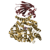
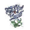
- PDB-1g2w: E177S MUTANT OF THE PYRIDOXAL-5'-PHOSPHATE ENZYME D-AMINO ACID AM... -
+ Open data

- Basic information
| Entry | Database: PDB / ID: 1g2w | ||||||
|---|---|---|---|---|---|---|---|
| Title | E177S MUTANT OF THE PYRIDOXAL-5'-PHOSPHATE ENZYME D-AMINO ACID AMINOTRANSFERASE | ||||||
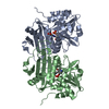
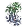
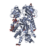
 Components Components | D-ALANINE AMINOTRANSFERASE | ||||||
 Keywords Keywords | TRANSFERASE / mutant / pyridoxal-5'-phosphate / water molecule / internal aldimine / aminotransferase / bacterial cell wall biosynthesis | ||||||
| Function / homology |  Function and homology information Function and homology informationD-amino acid biosynthetic process / D-amino-acid transaminase / D-alanine:2-oxoglutarate transaminase activity / D-amino acid catabolic process / pyridoxal phosphate binding / cytosol Similarity search - Function | ||||||
| Biological species |   Geobacillus stearothermophilus (bacteria) Geobacillus stearothermophilus (bacteria) | ||||||
| Method |  X-RAY DIFFRACTION / Resolution: 2 Å X-RAY DIFFRACTION / Resolution: 2 Å | ||||||
 Authors Authors | Lepore, B.W. / Ringe, D. | ||||||
 Citation Citation | Journal: Biochemistry and Molecular Biology of Vitamin B6 and PQQ-dependent Proteins, 10th Annual International Symposium on Vitamin B6 Year: 2000 Title: Studies on an Active Site Residue, E177, That Affects Binding of the Coenzyme in D-Amino Acid Transaminase, and Mechanistic Studies on a Suicide Substrate Authors: van Ophem, P.W. / Lepore, B.W. / Kishimoto, K. / Ringe, D. / Manning, J.M. #1: Journal: Biochemistry / Year: 1995Title: Crystal Structure of a D-amino Acid Aminotransferase: How the Protein Controls Stereoselectivity Authors: Sugio, S. / Petsko, G.A. / Manning, J.M. / Soda, K. / Ringe, D. #2: Journal: Biochemistry / Year: 1999Title: Effects of the E177K mutation in D-amino acid aminotransaminase. Studies on an essential coenzyme anchoring group that contributes to stereochemical fidelity Authors: van Ophem, P.W. / Peisach, D. / Erickson, S.D. / Soda, K. / Ringe, D. / Manning, J.M. | ||||||
| History |
|
- Structure visualization
| Structure viewer | Molecule: MolmilJmol/JSmol |
|---|
- Downloads & links
-Download
| PDBx/mmCIF format | 1g2w.cif.gz | 121.3 KB | Display | PDBx/mmCIF format |
|---|---|---|---|---|
| PDB format | pdb1g2w.ent.gz | 100 KB | Display | PDB format |
| PDBx/mmJSON format | 1g2w.json.gz | Tree view | PDBx/mmJSON format | |
| Others |  Other downloads Other downloads |
-Validation report
| Arichive directory | https://data.pdbj.org/pub/pdb/validation_reports/g2/1g2wftp://data.pdbj.org/pub/pdb/validation_reports/g2/1g2w | HTTPS FTP |
|---|
-Related structure data
| Related structure data | |
|---|---|
| Similar structure data |


-Links
 PDBj
PDBj
- Assembly
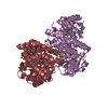
| Deposited unit | 
| ||||||||
|---|---|---|---|---|---|---|---|---|---|
| 1 |
| ||||||||
| Unit cell |
|
-Components
| #1: Protein | Mass: 32269.873 Da / Num. of mol.: 2 / Mutation: E177S Source method: isolated from a genetically manipulated source Source: (gene. exp.) Geobacillus stearothermophilus (bacteria)Plasmid: PICT113 / Production host: References: UniProt: P83771, UniProt: P19938*PLUS, D-amino-acid transaminase #2: Chemical |   Mass: 59.044 Da / Num. of mol.: 2 / Source method: obtained synthetically / Formula: C2H3O2 Mass: 59.044 Da / Num. of mol.: 2 / Source method: obtained synthetically / Formula: C2H3O2#3: Chemical |   Mass: 247.142 Da / Num. of mol.: 2 / Source method: obtained synthetically / Formula: C8H10NO6P Mass: 247.142 Da / Num. of mol.: 2 / Source method: obtained synthetically / Formula: C8H10NO6P#4: Water | ChemComp-HOH / |  Mass: 18.015 Da / Num. of mol.: 205 / Source method: isolated from a natural source / Formula: H2O Mass: 18.015 Da / Num. of mol.: 205 / Source method: isolated from a natural source / Formula: H2O |
|---|
-Experimental details
-Experiment
| Experiment | Method: X-RAY DIFFRACTION / Number of used crystals: 1 |
|---|
- Sample preparation
| Crystal | Density Matthews: 2.58 Å3/Da / Density % sol: 52.4 % | ||||||||||||||||||||||||||||||||||||||||||||||||||||||||||||
|---|---|---|---|---|---|---|---|---|---|---|---|---|---|---|---|---|---|---|---|---|---|---|---|---|---|---|---|---|---|---|---|---|---|---|---|---|---|---|---|---|---|---|---|---|---|---|---|---|---|---|---|---|---|---|---|---|---|---|---|---|---|
| Crystal grow | Temperature: 277 K / Method: vapor diffusion, hanging drop / pH: 8.5 Details: resevoir: Tris-HCl, pH 8.5, 22-26% PEG 4K, 0.2-0.4M NaOAc, 1mM alpha-ketoglutarate, enzyme buffer: 50mM KPO4, pH 7.2, 200mM KCl, 1mM DTT, 10uM PLP , VAPOR DIFFUSION, HANGING DROP, temperature 277K | ||||||||||||||||||||||||||||||||||||||||||||||||||||||||||||
| Crystal grow | *PLUS pH: 7.3 | ||||||||||||||||||||||||||||||||||||||||||||||||||||||||||||
| Components of the solutions | *PLUS
|
-Data collection
| Diffraction | Mean temperature: 277 K |
|---|---|
| Diffraction source | Source: ROTATING ANODE / Type: RIGAKU RU200 / Wavelength: 1.5418 |
| Detector | Type: RIGAKU RAXIS IIC / Detector: IMAGE PLATE / Date: Apr 29, 1999 |
| Radiation | Protocol: SINGLE WAVELENGTH / Monochromatic (M) / Laue (L): M / Scattering type: x-ray |
| Radiation wavelength | Wavelength: 1.5418 Å / Relative weight: 1 |
| Reflection | Resolution: 1.8→27 Å / Num. all: 303396 / Num. obs: 96070 / % possible obs: 86 % / Observed criterion σ(F): 1 / Observed criterion σ(I): -3 / Redundancy: 3.2 % / Biso Wilson estimate: 27.3 Å2 / Rmerge(I) obs: 0.123 / Net I/σ(I): 10 |
| Reflection shell | Resolution: 2.05→2.14 Å / Redundancy: 3.5 % / Rmerge(I) obs: 0.34 / Num. unique all: 1974 / % possible all: 92 |
| Reflection | *PLUS % possible obs: 85.4 % / Num. measured all: 303396 |
| Reflection shell | *PLUS Highest resolution: 1.8 Å / Lowest resolution: 1.86 Å / % possible obs: 97.5 % / Rmerge(I) obs: 0.244 |
- Processing
| Software |
| |||||||||||||||||||||||||
|---|---|---|---|---|---|---|---|---|---|---|---|---|---|---|---|---|---|---|---|---|---|---|---|---|---|---|
| Refinement | Resolution: 2→6 Å / Cross valid method: CNS free-R value test / σ(F): 1 / σ(I): -3 / Stereochemistry target values: Engh & Huber / Details: maximum likelihood target function on amplitudes
| |||||||||||||||||||||||||
| Solvent computation | Solvent model: CNS bulk solvent model | |||||||||||||||||||||||||
| Refinement step | Cycle: LAST / Resolution: 2→6 Å
| |||||||||||||||||||||||||
| Refine LS restraints |
| |||||||||||||||||||||||||
| LS refinement shell | Resolution: 2→2.12 Å / Total num. of bins used: 6
| |||||||||||||||||||||||||
| Software | *PLUS Name: CNS / Classification: refinement | |||||||||||||||||||||||||
| Refinement | *PLUS Highest resolution: 2 Å / Lowest resolution: 27 Å / σ(F): 1 / % reflection Rfree: 10 % | |||||||||||||||||||||||||
| Solvent computation | *PLUS | |||||||||||||||||||||||||
| Displacement parameters | *PLUS | |||||||||||||||||||||||||
| Refine LS restraints | *PLUS
| |||||||||||||||||||||||||
| LS refinement shell | *PLUS Highest resolution: 2 Å / Rfactor Rwork: 0.249 |
